Tomographic images from the interior of fossils, brain cells, or computer chips yield new insights into their finest structures. The 3-D images can be made with X-ray beams of the Swiss Light Source SLS, thanks to detectors and sophisticated computer algorithms the researchers developed themselves. PSI is a leader in nanotomography, which makes details visible on the scale of just a few millionths of a millimetre, and holds a world record for time-resolved tomography.
Fossils with a diameter of only half a millimetre are among the most exotic samples that have been examined at the large-scale research facility SLS. They had been discovered, by a British-Chinese team, in 609 million-year-old rocks in southern China. They were fossilised embryos from an organism that may have been a precursor to the first animals. Together with PSI researchers, the international team created three-dimensional images that showed details of less than a thousandth of a millimetre. The researchers recognised various stages of development and concluded that the organism reorganised its cells in the course of embryo development, just as humans and other animals living today do – an important step in evolution.
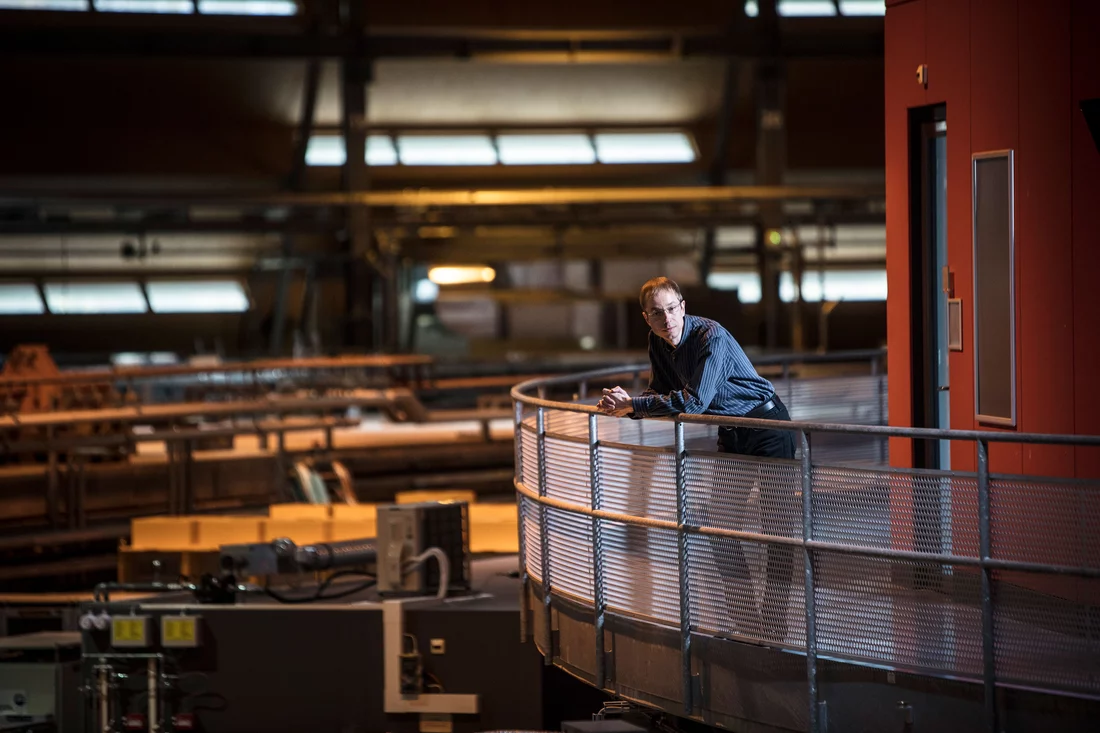
"We make tomographic images of the most diverse tissue and material samples, we look inside batteries, fuel cells, or ice cream, and we can watch, for example, how a 3-D printer makes an object out of plastic", explains Oliver Bunk, head of the Laboratory for Macromolecules and Bioimaging at PSI. The principle is the same as for computer tomography in a hospital. However, while in medicine the apparatus moves around the patient when taking the cross-sectional images, the sample is rotated at PSI, and the X-ray beam always strikes it from the same direction.
World record: More than 200 images per second
The X-ray light generated at SLS is much brighter and more concentrated than that of a medical X-ray system. This allows imaging of the smallest details and even tracking of dynamic processes. Thus a German team was able, together with PSI researchers, to watch the foaming of liquid aluminium as it happened. Metal foams are especially promising for lightweight construction. Thanks to a new measuring table that rotates extremely precisely and quickly, the group was able to document the foaming with 208 three-dimensional X-ray images per second, a world record in tomography.
The beamline that delivered the X-ray light for these time-resolved measurements was named TOMCAT. It is one of 17 beamlines used to conduct different experiments at SLS. With TOMCAT, 3-D images can be produced with a resolution of ten thousandths of a millimetre (0.1 micrometre). On another beamline called cSAXS, researchers can even penetrate into the millionth of a millimetre range. "Here we do nanotomography", Oliver Bunk explains. This requires a technique that was developed at PSI.
This method is called ptychography. "The name, which we all stumble over today, comes from the 1960s", Oliver Bunk explains. In those days, the German physicist Walter Hoppe had the idea for the technique, which could not yet be realised in the absence of modern computer technology. What is also needed is a light source with a characteristic that is familiar from laser light: Fixed temporal and spatial intervals are maintained between the individual light particles as they travel together. Experts refer to this as coherent light. SLS is not in fact a laser, but some of the X-rays generated are in this kind of unison. At PSI it was shown in 2007 that ptychography works with X-rays, and since then the method has been continuously improved.
Imaging with diffraction patterns instead of shadows
The method is partly similar to normal tomography in that the sample is scanned by the X-ray beam. Yet while the medical X-ray image corresponds to a shadow that is cast, ptychography creates a diffraction image of the illuminated area – a pattern of points with differing intensity that initially bears no resemblance to the sample. It is only with computer algorithms that the desired image can be calculated from hundreds of overlapping diffraction images. In order for the process to work, powerful detectors – X-ray cameras such as those developed at PSI – are required. These are produced today by the Swiss company Dectris, a PSI spin-off, which supplies them to synchrotron facilities worldwide.
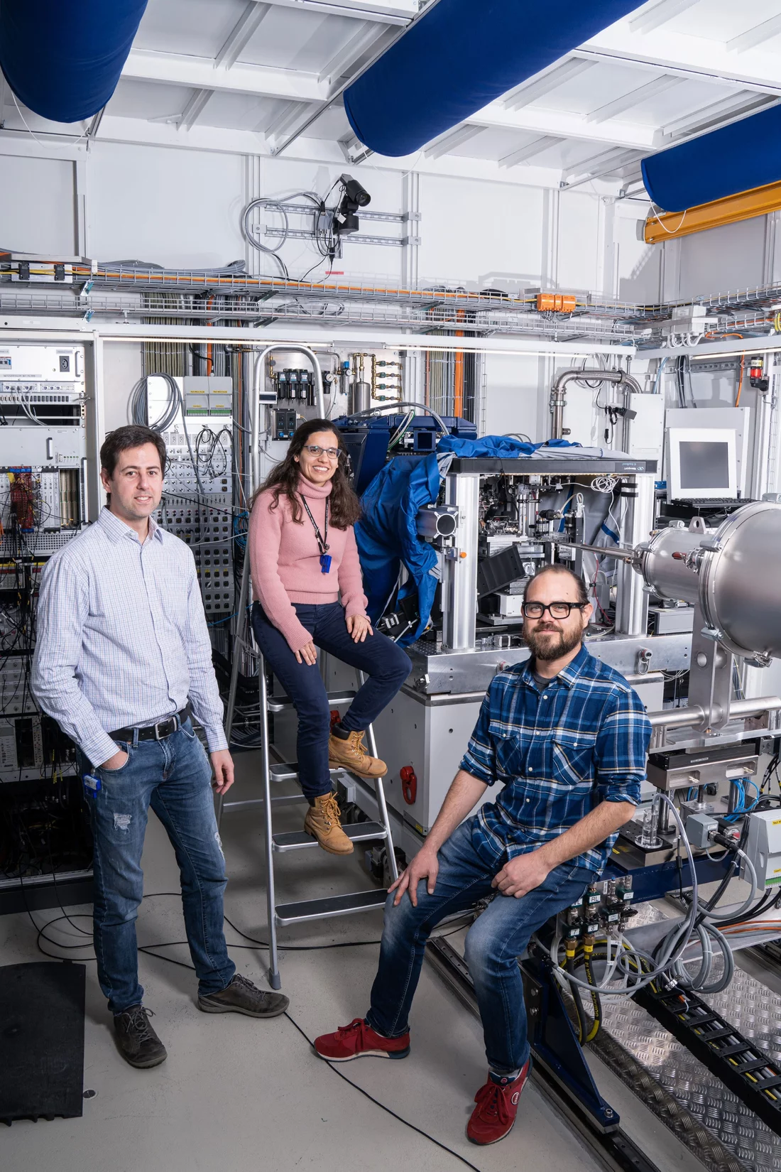
"We were the leaders in nanotomography, and we still are", says Oliver Bunk. The instruments built under the direction of PSI physicist Mirko Holler are unique worldwide. His colleague Manuel Guizar-Sicairos developed the algorithms for image reconstruction and received a prestigious international optics award in 2019. PSI physicist Ana Diaz provides the scientific background as an expert in biological tissue samples and material-physical issues.
Brain cells are among the particularly interesting samples that can be examined with the help of ptychography. In this way researchers would like to hunt down diseases such as Alzheimer's and Parkinson's. Three-dimensional images of bone structures have already yielded clues for osteoporosis research. For examination, tissue samples are deep-frozen and placed very precisely in a measuring chamber. For imaging in the nanometre range, the positional accuracy must be in this range too – "a big challenge", says Oliver Bunk: "A sample that is gigantic for us is about twice as thick as a human hair; small samples are ten times finer than the diameter of a hair."
X-raying catalysts and chips
Catalysts too are being investigated. They accelerate chemical processes and are indispensable in today's industry. "You would prefer to study catalysts while they're working", says Oliver Bunk. This is done at PSI using a variety of techniques, including ptychography. This enabled researchers to demonstrate what happens when a catalytic converter is subject to aging processes over the course of its operating life. The structural changes observed provide information about the processes taking place. This can be used, for example, to track when and how pores in a catalytic converter become clogged, or how the chemically active surfaces change their structure and thereby diminish the catalytic effect.
The PSI researchers are especially proud of their newest instrument, which bears the name LamNI. With it they can produce 3-D images of flat but rather expansive samples. "That is a big advance", comments Oliver Bunk. One of the first objects examined in this way was a computer chip. Earlier, the researchers had already X-rayed a commercially available chip and made the tiny transistors visible. But to do this, they had to cut out a small, cylindrical sample from the chip. With the new instrument, this is no longer necessary; the chip remains intact. "We can create an overview image and then zoom in, as you do with a camera, to make a high-resolution measurement", the physicist explains.
The 3-D images have a resolution of just under 20 nanometres (millionths of a millimetre) and can reveal if a chip is defective or even if it has been manipulated. The method could be used in high-security areas, such as power plants, where well protected IT hardware should be used to ensure that no unauthorised access is possible. The method could also be used for random sampling in quality control.
Oliver Bunk is convinced that nanotomography will be used even more widely in the future, because a new generation of synchrotron light sources provides even better conditions for this method. A corresponding upgrade is also planned for SLS. After this modernisation, the facility will deliver an even finer and more intense X-ray beam and thus significantly more coherent light. This will make it possible to carry out measurements more rapidly, or to reveal even smaller details in 3-D. The technologies required for this, such as better detectors, have already been developed at PSI and will be available in good time for measurements after the upgrade. With Swiss precision, the various scientific and high-tech developments mesh as tightly as gears to enable advances in many areas of fundamental and applied research.
Text: Barbara Vonarburg
Contact
Dr. Oliver Bunk
Laboratory for Macromolecules and Bioimaging
Paul Scherrer Institute, Forschungsstrasse 111, 5232 Villigen PSI, Switzerland
Telephone: +41 56 310 30 77, E-mail: oliver.bunk@psi.ch [German, English]


