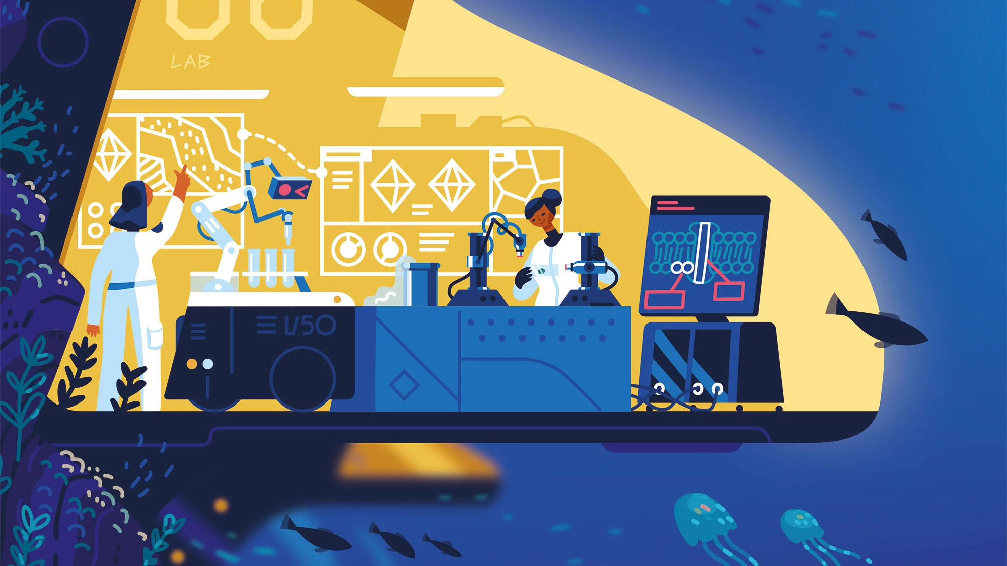
At PSI, researchers decipher the structure of the proteins in bacteria and viruses. This knowledge can aid, for example, in the development of drugs against infectious diseases. But before the investigation can begin, an extremely tricky problem has to be solved: the crystallisation of the molecules.
Chia-Ying Huang reaches for the glass cutter. Carefully yet forcefully, she cuts a small piece out of the thin glass plate in front of her. Now it becomes apparent that the plate, which initially looked like a single piece, has a thin film enclosed in the middle. Huang carves something out of it with a scalpel. Finally, with tweezers, she holds up a piece of film just a few millimetres wide. "My crystals are in here," she proudly proclaims.
To the naked eye, there's nothing there to see. Once Chia-Ying Huang puts the film under her microscope, though, even the most sceptical observer must admit that she's right: Dozens of colourless crystals appear on the edge of an enclosed drop of liquid. The translucent cubes look a bit like tiny diamonds. Huang clamps this precious snip of film in a holder and dips it into a bowl of liquid nitrogen. In there, other samples – preserved by the icy cold of the nitrogen – are already waiting to be measured at the Swiss Light Source SLS.
Biochemist Chia-Ying Huang has been working as a crystallographer at SLS for four years. Before that, she had developed a new method at Trinity College in Dublin, Ireland, to crystallise proteins in tiny amounts of liquid between two thin plastic films, in cooperation even then with researchers at SLS. Her colleagues at PSI and all external users who use SLS to decipher the structure of so-called membrane proteins now benefit from this technology. These biomolecules, which are naturally anchored in the cell wall of bacterial, animal, or human cells, are of great medical importance: A good third of all currently approved drugs act on this type of protein. But one property of these membrane proteins makes it especially difficult to crystallise them: They are not soluble in water. That's why you need particularly ingenious crystallisation methods to deal with them.
So small, and so coveted
The effort is worthwhile, because in order to explore the function of proteins and, for example, to develop drugs against viruses or bacteria, researchers need to know the structure of the proteins precisely. The most reliable way to find that out is crystal structure analysis. For this, the crystals are shot with synchrotron light; the structure of the molecules can be determined from the resulting diffraction pattern using, among other things, complex computational methods. For this to work, however, the protein molecules have to be arranged in a regular three-dimensional pattern: in other words, a crystal.
"Protein crystallisation is the bottleneck in structural biology," says May Sharpe. The crystallographer heads the PSI Crystallisation Facility, a unit that helps internal and external users of SLS grow the crystals they need for their research. "At the same time, there is something almost magical about this branch of science." There is no all-purpose formula for growing the crystals: Even after many decades of research, the experiments are still largely based on trial and error.
If you want to grow crystals of table salt or sugar, just dissolve a large amount of it in water and let the solution stand for a while. As a rule, beautiful crystals begin to grow. It's not so easy with proteins, though: They are complex molecules with a complicated three-dimensional structure. It is only very hesitantly that they organise themselves into regular, still rather loose associations. "For that, they have to be very pure and very stable," Sharpe sums up. "We like to say our proteins have to be happy."
Membrane proteins such as those Chia-Ying Huang works with are particularly delicate. "They are only really stable in their native environment: in the cell membrane," explains Sharpe. It is a major challenge to obtain this type of protein from the cells in a pure enough form, without the membrane, to grow crystals with them.
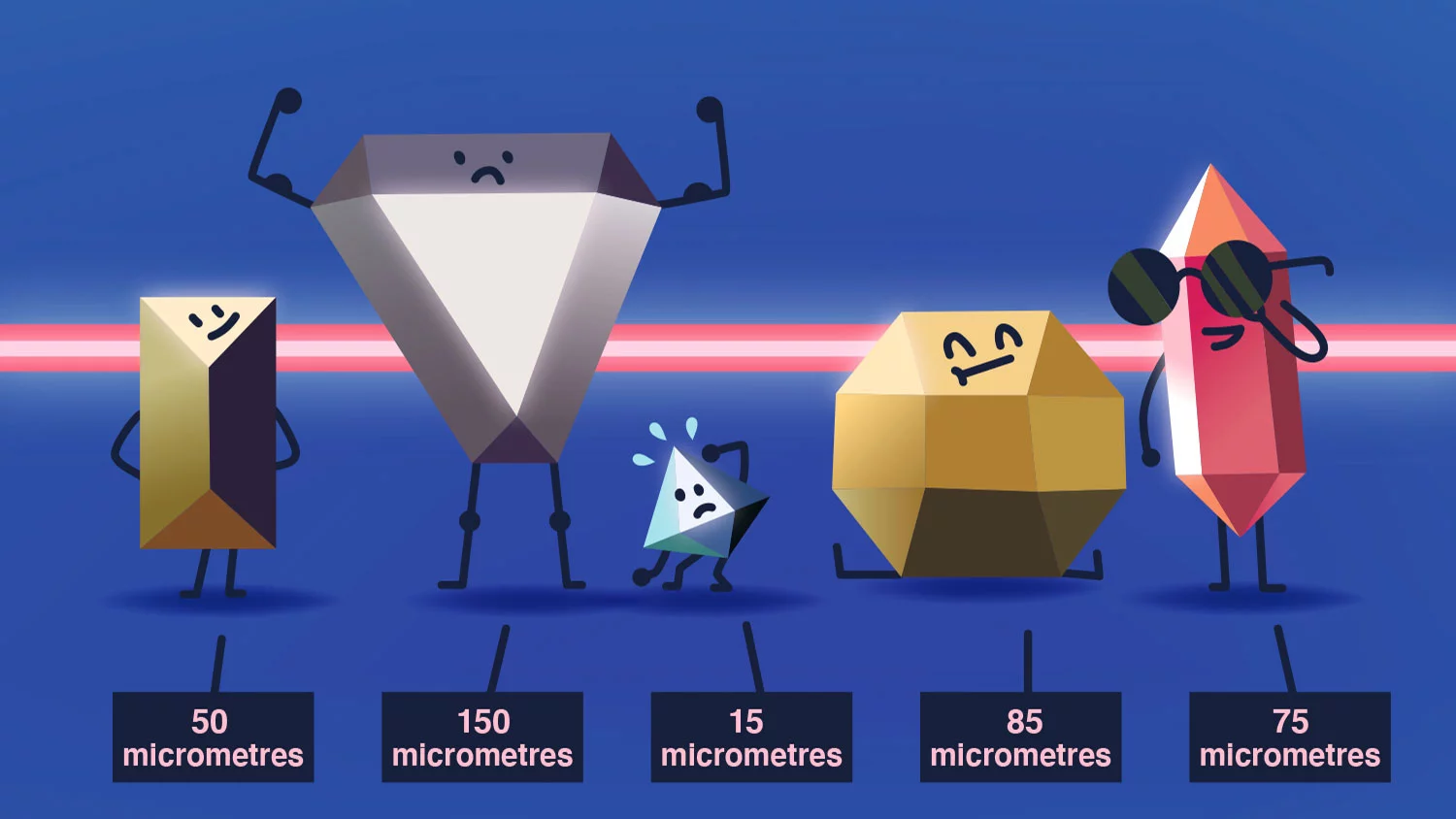
Protein crystals are tiny – especially in comparison to rock crystals you might see in a geology museum. With luck, they might be grown to a size of half a millimetre. Others remain so small that they can only be seen under the microscope.
A lot of persuasion is required
In Huang's laboratory at SLS, a pipetting robot slowly patrols a plastic plate with wells. Into each of the little wells, it pipettes tiny amounts of protein solution, just a ten-thousandth of a millilitre. Usually, the robot would then add a prepared mixture of water, salts, and chemical additives that have proven over time to be conducive to crystal formation. Since membrane proteins do not dissolve in water, Huang takes a different approach here. Either she adds an agent that stabilises the proteins, or she replaces water as a solvent with lipids, fat-loving biomolecules. In the method she developed herself, the robot pipettes the protein, together with lipids and auxiliary substances, between films in such a way that the film completely encloses the tiny lipid droplets. The liquid is safely stored in between; neither air nor moisture can interfere.
"With this method, the crystals of some types of membrane proteins grow much better," explains Chia-Ying Huang. "And there is another advantage: You don't have to fish them out of the solution once they have formed." Instead, Huang simply uses the glass cutter to remove the shrink-wrapped droplets, along with the crystals, from the plate.
Chia-Ying Huang's membrane proteins yield especially tiny crystals with an edge length of only a tenth or even a hundredth of a millimetre. "Such small crystals are easily destroyed in the SLS X-ray beam," she explains. "So we have to measure many of them one after the other to get enough data." Fortunately, when everything goes well, several hundred crystals per sample are usually formed. One after the other, they are scanned and measured by the SLS X-ray beam. This is made possible by so-called serial crystallography.
In the plate hotel, there are a thousand beds
The heart of the Crystallisation Facility at PSI is the "plate hotel”, as the researchers call this large laboratory cabinet, which is kept at a temperature of 20 degrees Celsius. In it are stored up to a thousand plastic plates with dozens of samples each. The researchers wait for something to happen in the protein solutions – more precisely, for crystals to precipitate. The plates are regularly moved automatically under a camera, which photographs each individual well and stores the photo.
Chia-Ying Huang looks at images from one such series of experiments on the computer screen. "The solutions have been in there for three weeks now, but you still can't see anything," she says, pointing to the countless photos of colourless drops. But this doesn't frustrate her. "It usually takes a month for crystals to form." Quite often, however, nothing happens. The crystallographer then uses the pipetting robot to prepare new protein solutions with a different composition: She increases the protein concentration, changes the solvent, or introduces additives – and then waits some more.
The proteins don't always put the researchers' patience to the test. On the contrary: Some precipitate in a big, disorderly heap. The quality of the solid substances formed in this way is not adequate for crystal structure analysis, however, since the molecules are not arranged in regular patterns. Here, too, it means: Try again – maybe this time with a dilute solution.
Basically, you're just staring at drops.
"Protein crystallisation is a thankless job," says May Sharpe with a laugh. "Basically, you're just staring at drops. Most researchers don't hate it, but I like it just fine." She developed her passion for this field during her doctoral research at the biotechnology and pharmaceutical company Novartis in Basel, where she explored the crystallisation of proteins for the purpose of drug development. What her colleagues saw as a necessary but unpleasant step quickly aroused her interest.
"I enjoy trying so many different things. And it's nice to help others get hold of these coveted crystals," she says. What has May Sharpe learned about growing protein crystals in her eight years at PSI? "Don’t think too much," she says. "Instead, just try, look and learn. Above all, however, you have to get rid of the idea that you always understand everything that is going on." This is exactly what constitutes the almost magical component of her work when diving deep into the world of microbes.
Text: Brigitte Osterath
Copyright
PSI provides image and/or video material free of charge for media coverage of the content of the above text. Use of this material for other purposes is not permitted. This also includes the transfer of the image and video material into databases as well as sale by third parties.


