Show filters
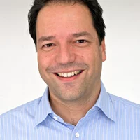
Brand new I-TOMCAT beamline comes to life
Marking another milestone in the TOMCAT 2.0 upgrade project, I-TOMCAT — our newly built beamline — received its first X-ray light on September 25, 2025.

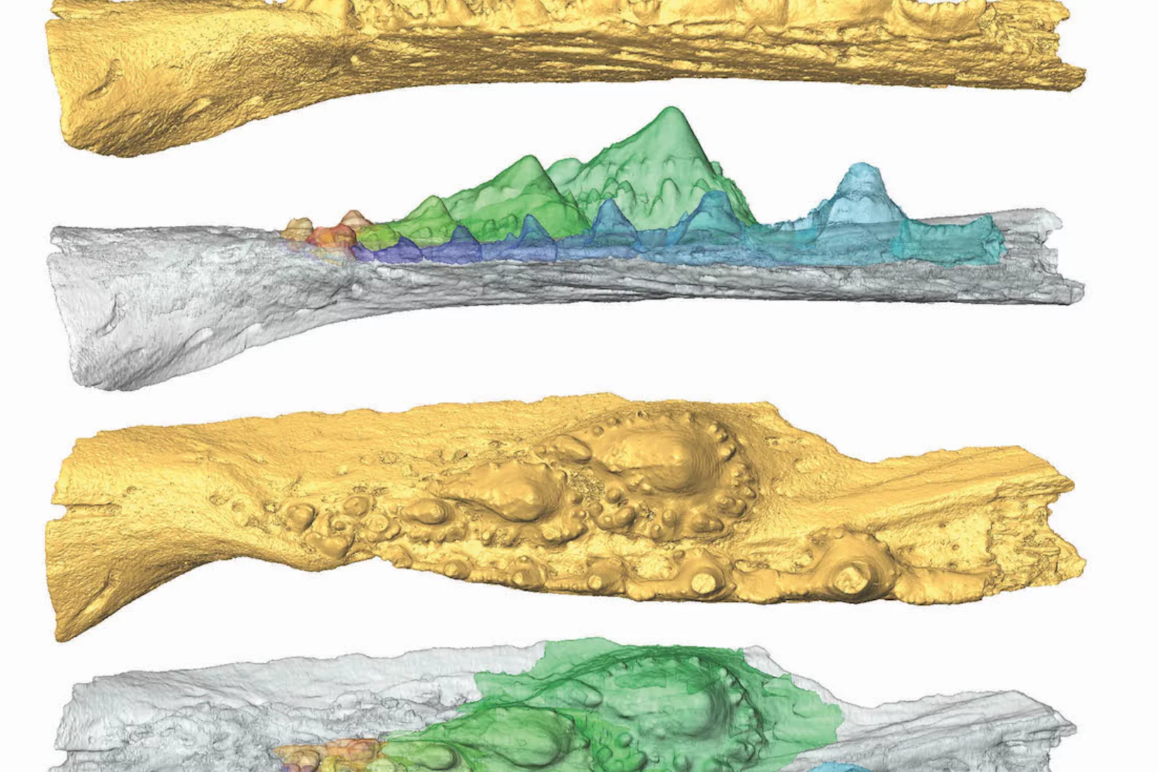
Big heart, acute senses key to explosive radiation of early fishes
X-rays of a 400-million-year-old fossil illuminate a key moment in our deep evolutionary past.

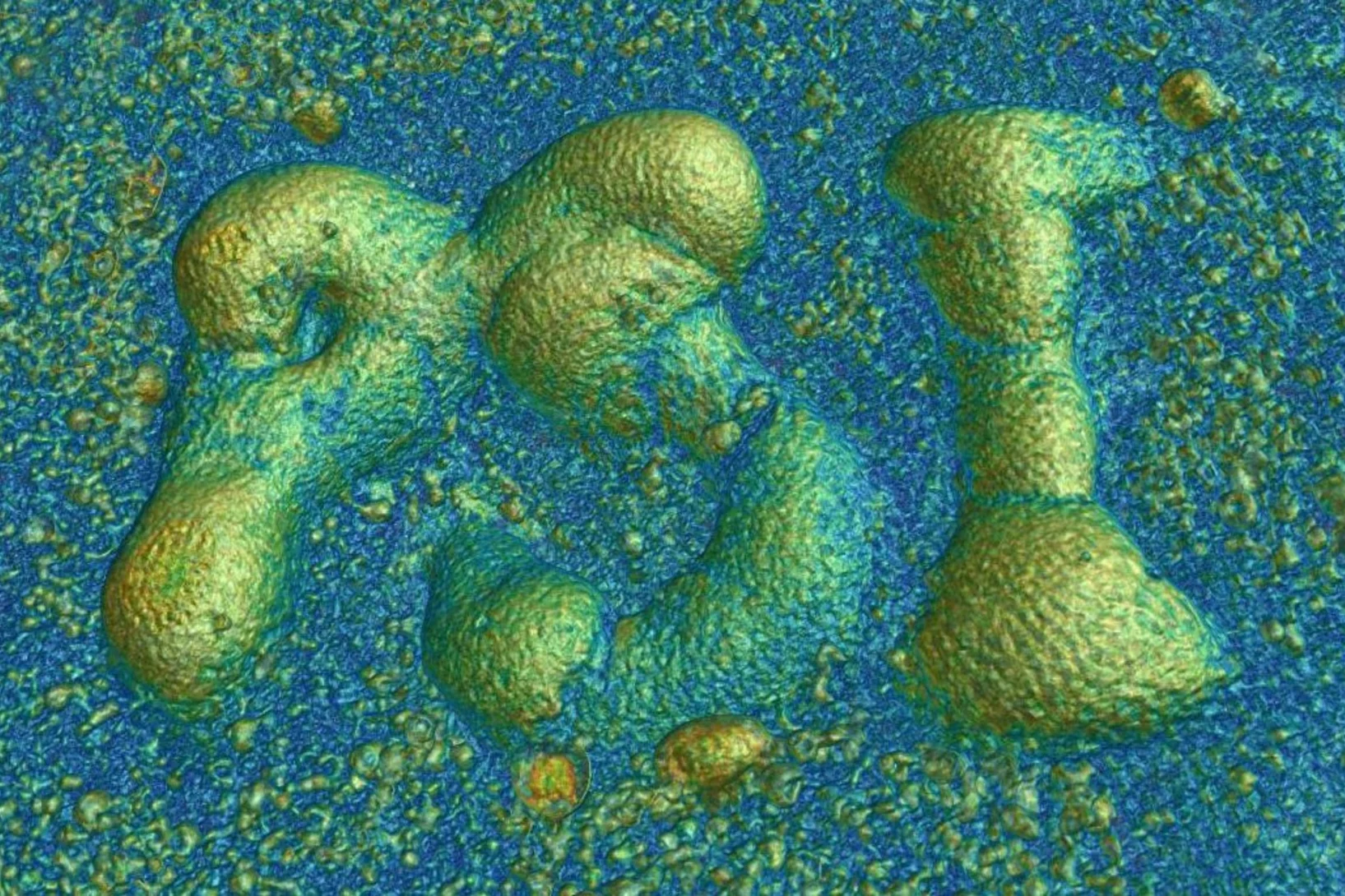
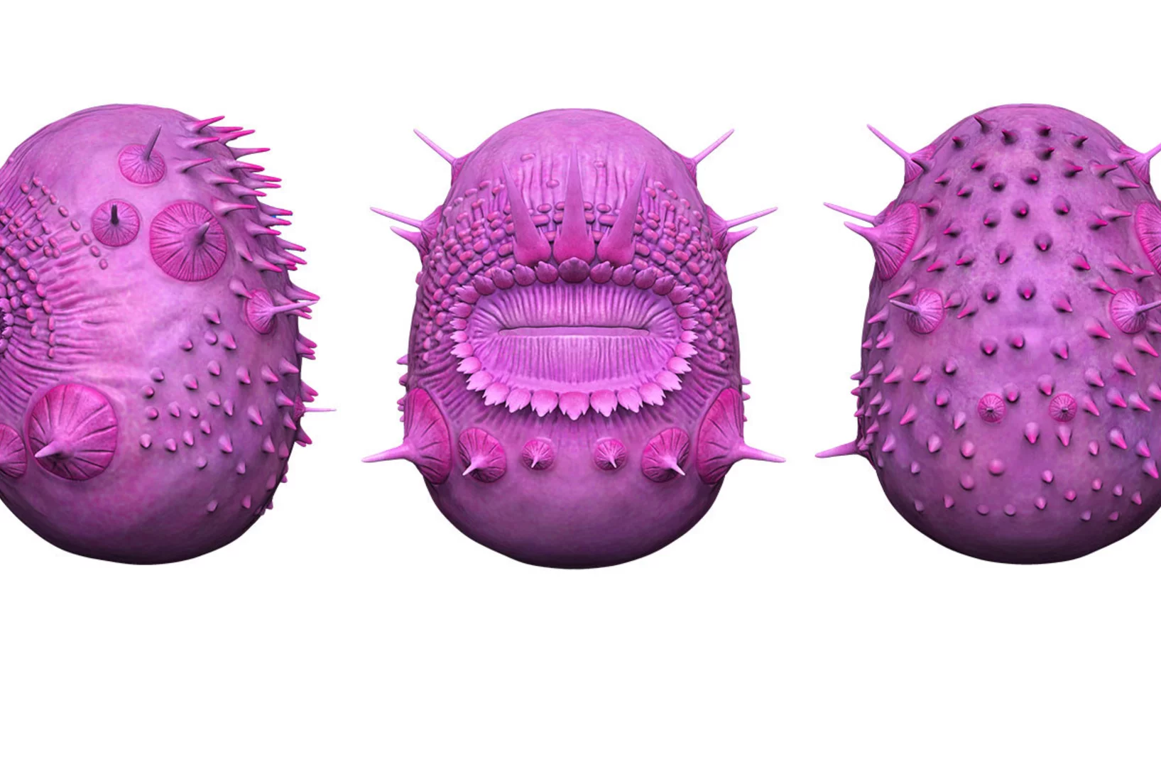
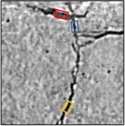
X-rays reveal fossil stealth technology
PSI imaging helps to uncover the hunting strategy of a prehistoric predator.
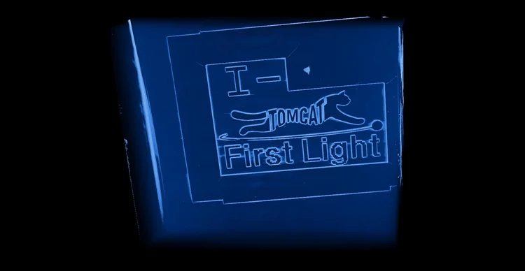
Zinc detected in clogged syringes
With the help of researchers at PSI, ANAXAM has been investigating, on behalf of the pharmaceutical company MSD, whether zinc may contribute to clogging of pre-filled syringes.
S-TOMCAT beamline receives first X-ray light
On June 25th 2025, the S-TOMCAT beamline shutters were opened to receive first X-ray light from the new SLS 2.0 storage ring.
Cause of clogged hypodermic needles discovered
Researchers at PSI and the ANAXAM technology transfer center have found the cause of clogging in prefilled syringes.

Listening for Defects as They Happen
Experiments at the Swiss Light Source SLS help resolve a long-standing debate surrounding metal 3D laser printing.
3D insights into an innovative manufacturing process
3D printing for creating complex shapes
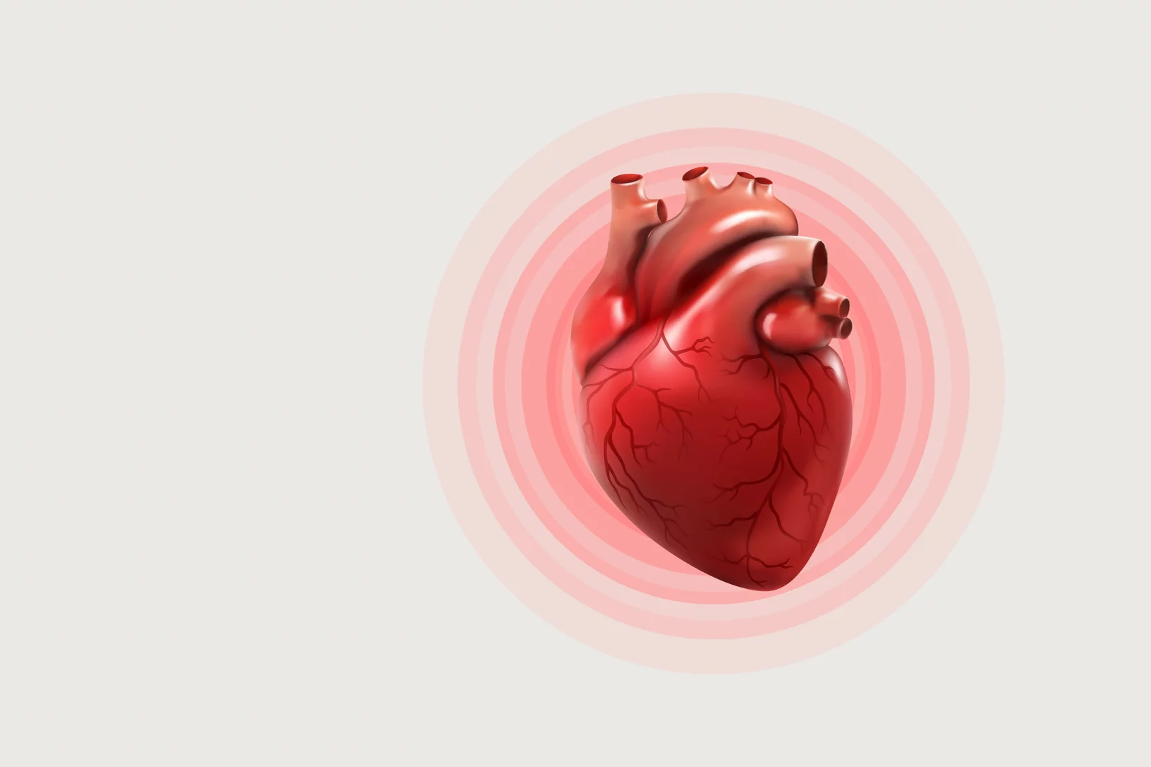
X-ray imaging after heart transplantations
Synchrotron light can be used in follow-up after a heart transplant to determine whether the body may be rejecting the new organ.
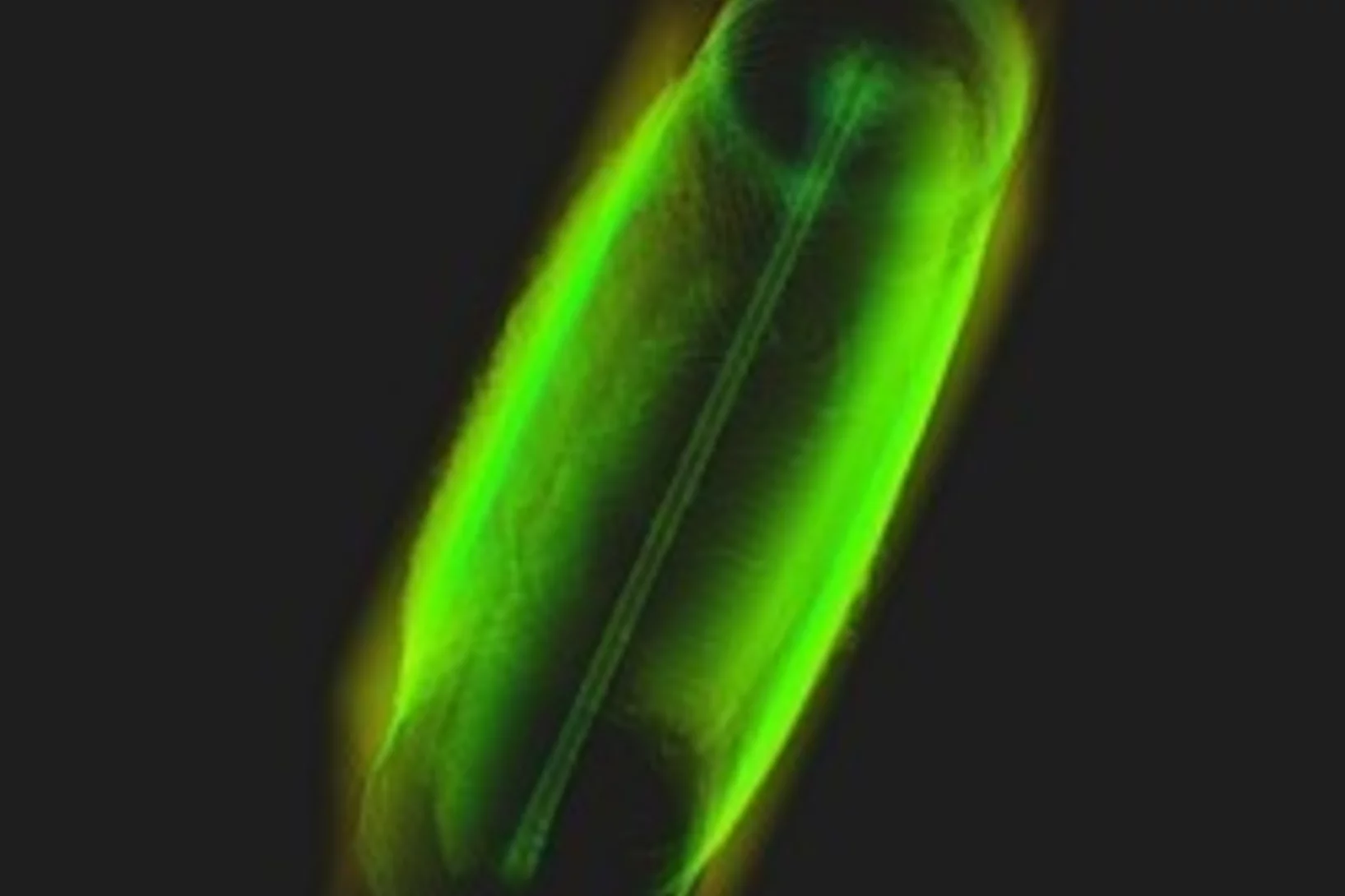
X-ray tomography helps understand how the heart beats
Researchers at the Swiss Light Source SLS use X-ray phase contrast imaging to study a heart in action as it beats.

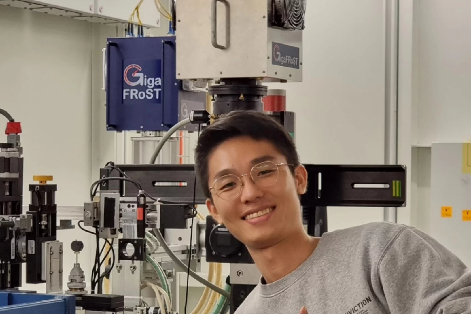
Jisoo Kim receives PSI Thesis Medal 2023
Jisoo Kim receives the PSI Thesis Medal 2023. With this award, PSI recognises outstanding PhD theses, achieving a high degree of innovation and potentially leading to scientific breakthroughs. Jisoo holds a Master of Science from the Korean Advanced Institute of Science &Technology and defended his thesis entitled “Towards time-resolved X-ray scattering tensor tomography” at ETH Zürich.
Weird fossil is not our ancestor
X-ray light solves puzzle of human ancestry
Jisoo Kim bags the 2022 Werner Meyer-Ilse Award
Jisoo Kim was awarded the 2022 Werner Meyer-Ilse Memorial Award. The WMI Award is given to young scientists for exceptional contributions to the advancement of X-ray microscopy through either outstanding technical developments or applications, as evidenced by their presentation at the International Conference on X-ray Microscopy and supporting publications. Jisoo was awarded for his development of the method "Time-resolved x-ray scattering tomography for rheological studies", and is co-recipient of the award with Yanqi Luo from the Advanced Photons Source for her work on applications. The award was presented during the 15th International Conference on X-ray Microscopy XRM2022 hosted by the National Synchrotron Radiation Research Center (NSRRC) in Hsinchu, Taiwan on 19 - 24 June, 2022.
Direct observation of crack formation mechanisms with operando Laser Powder Bed Fusion X-ray radiography
Operando high-speed X-ray radiography experiments reveal the cracking mechanism during 3D laser printing of a Ni superalloy.
X-ray microscopy with 1000 tomograms per second
A team at the Swiss Light Source SLS have set a new record using an imaging method called tomoscopy.

Hierarchical imaging and computational analysis of three-dimensional vascular network architecture in mouse brain
An international team involving researchers from the University and University Hospital Zürich, the Krembil Research Institute and the University and University Hospital in Toronto (Canada), the Department of Physics of Jyväskylä (Finland), the University of Leuven (Belgium), the Johannes Kepler University in Linz (Austria), the Novartis Institutes for Biomedical Research in Emeryville (USA), the ETH Zürich and the Paul Scherrer Institute has developed a protocol that enables hierarchical imaging and computational analysis of vascular networks in entire postnatal- and adult mouse brains, enabling direct and quantitative comparisons of the morphological brain vascular network architecture between different postnatal and / or adult developmental stages. The results have been published on Nature Protocols on September 3rd, 2021.
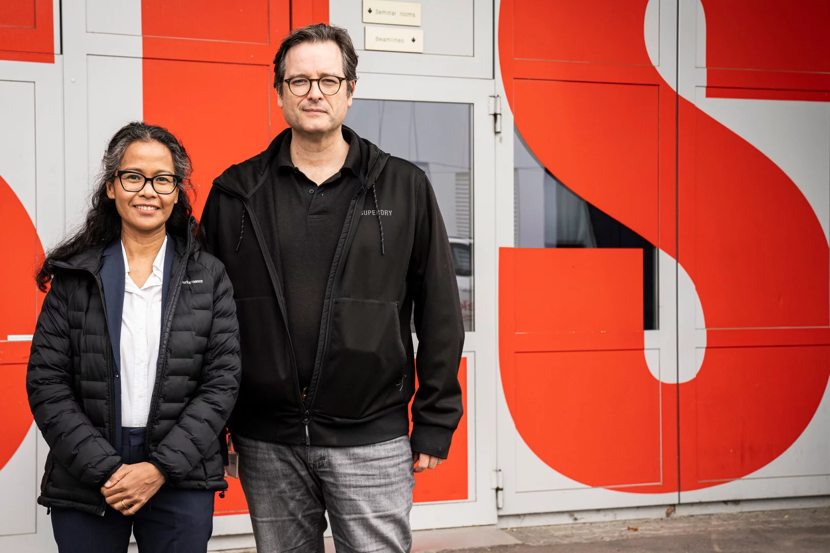
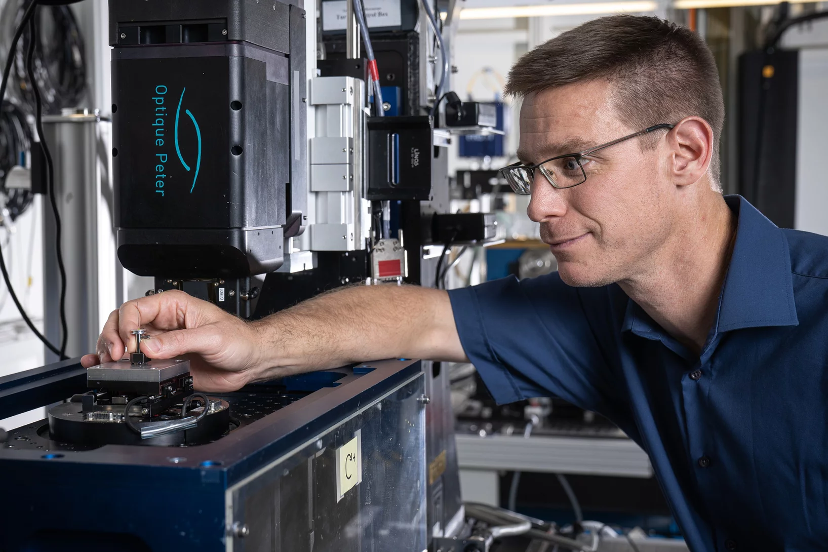
TOMCAT welcomes on board two scientists
The X-ray Tomography group welcomes on board Mariana Verezhak and Goran Lovric as members of the TOMCAT beamline crew. They will both contribute to the further development and realization of TOMCAT 2.0 (S- and I-TOMCAT branches on SLS2.0).
Understanding Why Solid-State Batteries Fail
Researchers from the University of Oxford, the Diamond Light Source and the Paul Scherrer Institut have generated strong evidence supporting one of two competing theories regarding the mechanism by which lithium metal dendrites grow through ceramic electrolytes. A process leading to short circuit at high rates of charge. The X-ray phase-contrast imaging capabilities of the TOMCAT beamline of the Swiss light source allowed researchers to visualize and characterize the growth of cracks and dendrites deep within an operating solid-state battery. The results were published in Nature Materials on April 22, 2021.
Deep evolutionary origins of the human smile
Detailed characterization of the tooth and jaw structure and development among shark ancestors by synchrotron based X-ray tomographic microscopy at TOMCAT led an international team of researchers from the Naturalis Biodiversity Center in Leiden and the University of Bristol to the discovery that while teeth evolved once, complex dentitions have been gained and lost many times in evolutionary history.

SLS 2.0 approved - TOMCAT 2.0 cleared for takeoff!
In December 2020 the Swiss parliament approved the Swiss Dispatch on Promotion of Education, Research and Innovation (ERI) for 2021 to 2024 which includes funding for the planned SLS 2.0 upgrade. The new machine will lead to significantly increased brightness, thus providing a firm basis for keeping the SLS and its beamlines state-of-the-art for the decades to come. The TOMCAT crew is very excited that the TOMCAT 2.0 plans (deployment of the S- and I-TOMCAT branches, see SLS 2.0 CDR, p. 353ff) have been included in the Phase-I beamline upgrade portfolio. These beamlines will receive first light right after the commissioning of the SLS 2.0 machine around mid 2025. A first milestone towards this goal has just been achieved, with the successful installation of the S-TOMCAT optics hutch during W1 of 2021. The TOMCAT scientific and technical staff would like to thank Mr. Nolte and his Innospec crew for delivering perfectly on schedule.

BEATS beamline scientist from SESAME synchrotron trains at TOMCAT
TOMCAT welcomes Gianluca Iori, beamline scientist from BEATS - the new beamline for tomography at the SESAME synchrotron in Jordan, to a 3-month training on beamline operations. Gianluca’s visit is part of the Staff Training (BEATS Work Package 2) organized for BEATS scientific staff and SESAME control engineers. BEATS is a European project, funded under the EU’s Horizon 2020 research and innovation programme and coordinated by the ESRF.
3 new Post Docs and 1 PhD student join TOMCAT
The X-ray Tomography group welcomes Stefan Gstöhl (Post-Doc), Maxim Polikarpov (Post-Doc), Margaux Schmeltz (Post-Doc) and Aleksandra Ivanovic (PhD Student) as new members. The group also thank everybody who helped making it possible for our Post-Docs and PhD student to join PSI amidst the challenges brought by the COVID-19 pandemic.

Automatic extraction of dynamic features from sub-second tomographic microscopy data
A fully automatized iterative reconstruction pipeline designed to reconstruct and segment dynamic processes within a static matrix has been developed at TOMCAT. The algorithm performance is demonstrated on dynamic fuel cell data where it enabled automatic extraction of liquid water dynamics from sub-second tomographic microscopy data. The work is published in Scientific Reports on 2 October 2020.

3D printing silica aerogels at the micrometer scale
A group of EMPA and ETH Zürich researchers have developed a new method to directly write ink made of silica aerogels in 3D. Thanks to X-ray phase contrast tomography at the TOMCAT beamline they characterized the resulting printed material with different compositions. Their results were published in Nature on August 18, 2020.
")
Phase contrast microtomography reveals nanoparticle accumulation in zebrafish
Metal-based nanoparticles are a promising tool in medicine – as a contrast agent, transporter of active substances, or to thermally kill tumor cells. Up to now, it has been hardly possible to study their distribution inside an organism. Researchers at the University of Basel in collaboration with the TOMCAT team have used phase contrast X-ray tomographic microscopy to take high-resolution captures of the nanoparticle aggregation inside zebrafish embryos.
The study was published in the journal Small and featured on the cover of its current issue.
4 times compression factor for tomographic data feasible
In a recent study, TOMCAT has shown that lossy compression by a factor of at least 3 to 4 of raw acquisitions generally does not affect the reconstruction quality and that higher factors (six to eight times) can be achieved for tomographic volumes with a high signal-to-noise ratio as it is the case for phase-retrieved datasets. This finding is relevant to current challenges on large tomography data management and storage especially at synchrotron facilities. The results of this study was published in Journal of Synchrotron Radiation.
Miniaturized fluidic circuitry observed in 3D
The team of Prof. Thomas Hermans at the University of Strasbourg in France managed to create wall-less aqueous liquid channels called anti-tubes. Thanks to X-ray phase contrast tomography at the TOMCAT beamline those anti-tubes could be observed in 3D. The exciting results were published in Nature on May 6, 2020.

Rapid 3D directional small-angle scattering imaging achieved at TOMCAT
Researchers from the TOMCAT beamline have developed a small-angle scattering tensor tomography method to visualize microscopic features within a macroscopic field of view with unprecedented data acquisition speed. The results of the study were published in Applied Physics Letters on April 1, 2020.

X-ray Imaging for Biomedicine: Imaging Large Volumes of Fresh Tissue at High Resolution
The TOMCAT beamline at the Swiss Light Source specializes in rapid high-resolution 3-dimensional tomographic microscopy measurements with a strong focus on biomedical imaging. The team has recently developed a technique to acquire micrometer-scale resolution datasets on the entire lung structure of a juvenile rat in its fresh natural state within the animal’s body and without the need for any fixation, staining or other alteration that would affect the observed structure (E. Borisova et al., 2020, Histochem Cell Biol).

Towards dynamic feedback control during time-resolved CT at TOMCAT
Researchers from the CWI in Amsterdam and the TOMCAT beamline have developed and implemented a real-time CT reconstruction, visualisation, and on-the-fly analysis approach to monitor dynamic processes as they occur. With processes of multiple sets of CT slices per second, this represents the next crucial step towards adaptive feedback control of time-resolved in situ tomographic experiments. The results of this study were published in Scientific Reports on December 5, 2019.