
Researchers at the Swiss Light Source SLS’s TOMCAT beamline have used phase contrast X-ray imaging to study a rat’s heart as it beats. The work will pave the way to investigations into how the heart beats on a cellular level, and understanding the effect of disease and drugs on the cardiac cycle.
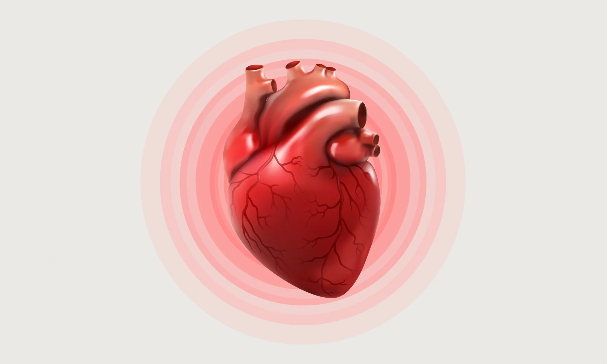
How the heart works may be the subject of a high school biology class. But how it really works on a cellular level is in fact not fully understood. The muscle cells that make the heart contract are called cardiomyocytes. Central to their function is their three dimensional arrangement in the muscular walls of the heart, which evolves throughout the cardiac cycle. As the heart beats, the cardiomyocytes aggregate and orientate themselves into fibres in such a way that determines the propagation of electrical currents and thus the force generated within the heart. However, how this process really takes place remains unknown, as most experimental techniques do not allow something as small as a cell to be evaluated while they function within the organ.
Here, in steps the Swiss Light Source's TOMCAT beamline with phase-contrast tomographic X-ray imaging. This technique gives the advantage of multiscale imaging: allowing something very small (the cellular structure) to be studied in something very big (the heart). Now, thanks to a specially designed perfused isolated heart system (‘Langendorff’), a team of researchers were able for the very first time to study the heart in action as it beats at an unprecedented resolution. “This system combined with the ultra-fast endstation that we have at the TOMCAT beamline allows us to make dynamic measurements, giving us unique access to how the heart is beating,” says TOMCAT beamline scientist Anne Bonnin, who leads the project in collaboration with colleagues in Bern and Barcelona.
Because you have access to how the cardiac fibres change over time, you can foresee different types of studies: for example, what happens if the heart has a stroke or if we ingest certain drugs
A Langendorff heart compatible with X-ray tomography
The Langendorff system keeps the heart beating after it has been removed from the circulatory system of the donor – in this case, a rat. This is achieved by perfusing the heart with nutrient rich, oxygenated buffer solutions under carefully controlled conditions. The team created a customised Langendorff system, notably that can rotate, which it is compatible with phase contrast tomographic X-ray imaging. Using their set up, the team were able to record the heart beating in three dimensions at an unprecedented resolution and observe the changing cardiomyocyte aggregates and other larger cardiac structures.

A high-resolution means to study how drugs and disease affect the cardiac cycle
Now that the feasibility of the system has been demonstrated, the work paves the way to high-resolution studies on the dynamics of the heart structure, including diseases or drugs that affect this, as Anne Bonnin explains. “Because you have access to how the cardiac fibres change over time, you can foresee different types of studies: for example, what happens if the heart has a stroke, how it differs with different heartrates or what happens if we ingest certain drugs.”
The SLS 2.0 upgrade, during which TOMCAT is set to receive a phase-I beamline upgrade in 2025, promises to be a game-changer for this work. At present, Bonnin comments, the field of view only covers a small part of the heart at a time. The higher coherence and flux of SLS 2.0 combined with the beamline upgrade will make it possible to improve the image quality drastically and achieve image of an entire heart cross section. Indeed, higher coherence will provide greater contrast – a crucial factor for the phase contrast X-ray imaging team, who rely on the phase changes of a coherent beam in different materials.
Text: Paul Scherrer Institute / Miriam Arrell
Original Publication
A tomographic microscopy-compatible Langendorff system for the dynamic structural characterization of the cardiac cycle
Hector Dejea, Christian M. Schlepütz, Natalia Méndez-Carmona, Maria Arnold, Patricia Garcia-Canadilla, Sarah L. Longnus, Marco Stampanoni, Bart Bijnens and Anne Bonnin
Frontiers in Cardiovascular Medicine, 22nd December 2022
DOI: 10.3389/fcvm.2022.1023483
Contact
Dr. Anne Bonnin
TOMCAT Beamline Scientist
Paul Scherrer Institute, Forschungsstrasse 111, 5232 Villigen PSI, Switzerland
Telephone: +41 56 310 46 78, e-mail: anne.bonnin@psi.ch
Further Information
Heart Imaging Project | X-Ray Tomography | Paul Scherrer Institut (PSI)
Copyright
PSI provides image and/or video material free of charge for media coverage of the content of the above text. Use of this material for other purposes is not permitted. This also includes the transfer of the image and video material into databases as well as sale by third parties.