
News
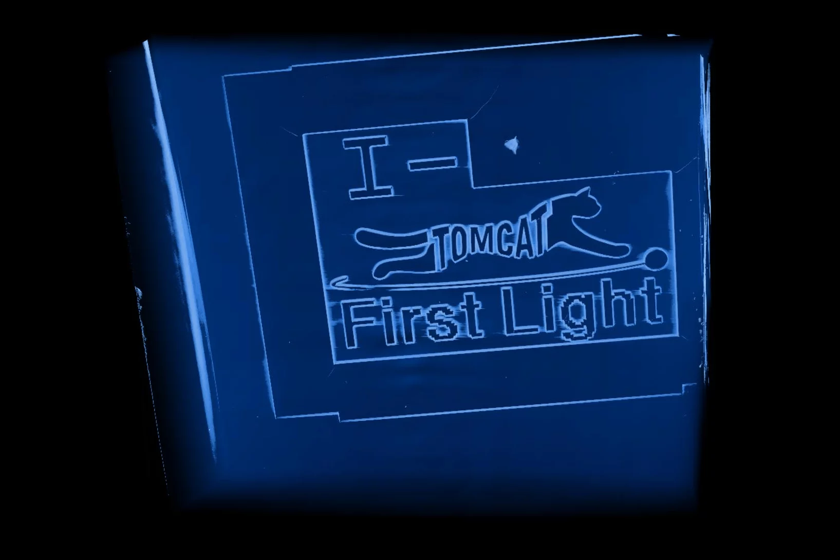
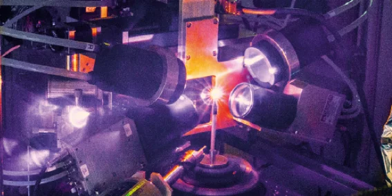
Brand new I-TOMCAT beamline comes to life
Marking another milestone in the TOMCAT 2.0 upgrade project, I-TOMCAT — our newly built beamline — received its first X-ray light on September 25, 2025.
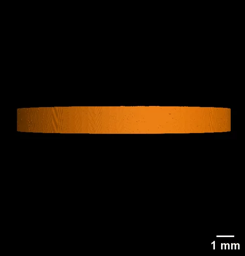
S-TOMCAT beamline receives first X-ray light
On June 25th 2025, the S-TOMCAT beamline shutters were opened to receive first X-ray light from the new SLS 2.0 storage ring.

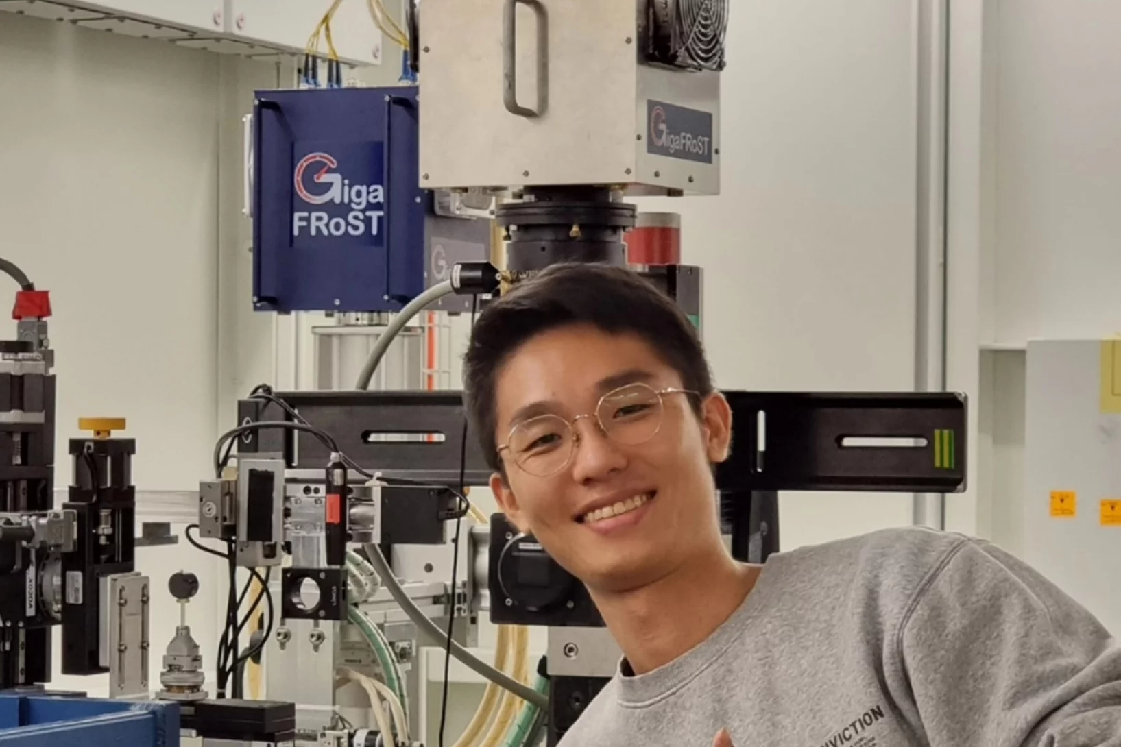
Jisoo Kim receives PSI Thesis Medal 2023
Jisoo Kim receives the PSI Thesis Medal 2023. With this award, PSI recognises outstanding PhD theses, achieving a high degree of innovation and potentially leading to scientific breakthroughs. Jisoo holds a Master of Science from the Korean Advanced Institute of Science &Technology and defended his thesis entitled “Towards time-resolved X-ray scattering tensor tomography” at ETH Zürich.
Jisoo Kim bags the 2022 Werner Meyer-Ilse Award
Jisoo Kim was awarded the 2022 Werner Meyer-Ilse Memorial Award. The WMI Award is given to young scientists for exceptional contributions to the advancement of X-ray microscopy through either outstanding technical developments or applications, as evidenced by their presentation at the International Conference on X-ray Microscopy and supporting publications. Jisoo was awarded for his development of the method "Time-resolved x-ray scattering tomography for rheological studies", and is co-recipient of the award with Yanqi Luo from the Advanced Photons Source for her work on applications. The award was presented during the 15th International Conference on X-ray Microscopy XRM2022 hosted by the National Synchrotron Radiation Research Center (NSRRC) in Hsinchu, Taiwan on 19 - 24 June, 2022.
Scientific Highlights

Kräftiges Herz, hochentwickelte Sinne – der Schlüssel zur rasanten evolutionären Vielfalt früher Fische
Röntgenaufnahmen eines 400 Millionen Jahre alten Fossils beleuchten einen Schlüsselmoment in unserer frühen evolutionären Vergangenheit

Fossile Tarntechnologie im Röntgenlicht
PSI-Bildgebung hilft, die Jagdstrategie eines prähistorischen Urzeiträubers zu entschlüsseln.
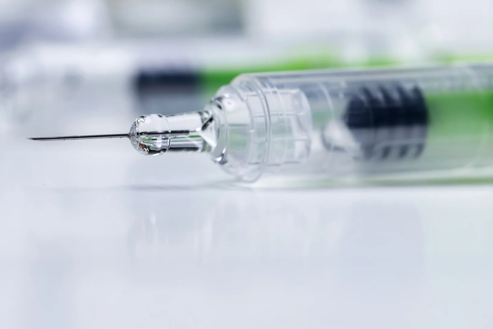
Zink in verstopften Spritzen nachgewiesen
Für das Pharmaunternehmen MSD hat ANAXAM mithilfe von Forschenden des PSI untersucht, ob Zink zur Verstopfung von Fertigspritzen beitragen kann.
Ursache für verstopfte Spritzennadeln gefunden
Forschende des PSI und des Technologietransferzentrums ANAXAM finden die Ursache für Verstopfungen bei vorgefüllten Fertigspritzen.