
News
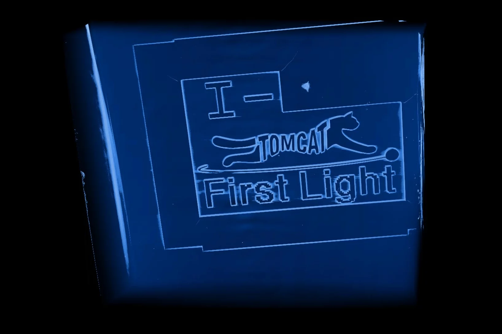
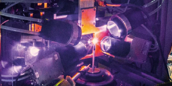
Brand new I-TOMCAT beamline comes to life
Marking another milestone in the TOMCAT 2.0 upgrade project, I-TOMCAT — our newly built beamline — received its first X-ray light on September 25, 2025.
S-TOMCAT beamline receives first X-ray light
On June 25th 2025, the S-TOMCAT beamline shutters were opened to receive first X-ray light from the new SLS 2.0 storage ring.

Jisoo Kim receives PSI Thesis Medal 2023
Jisoo Kim receives the PSI Thesis Medal 2023. With this award, PSI recognises outstanding PhD theses, achieving a high degree of innovation and potentially leading to scientific breakthroughs. Jisoo holds a Master of Science from the Korean Advanced Institute of Science &Technology and defended his thesis entitled “Towards time-resolved X-ray scattering tensor tomography” at ETH Zürich.
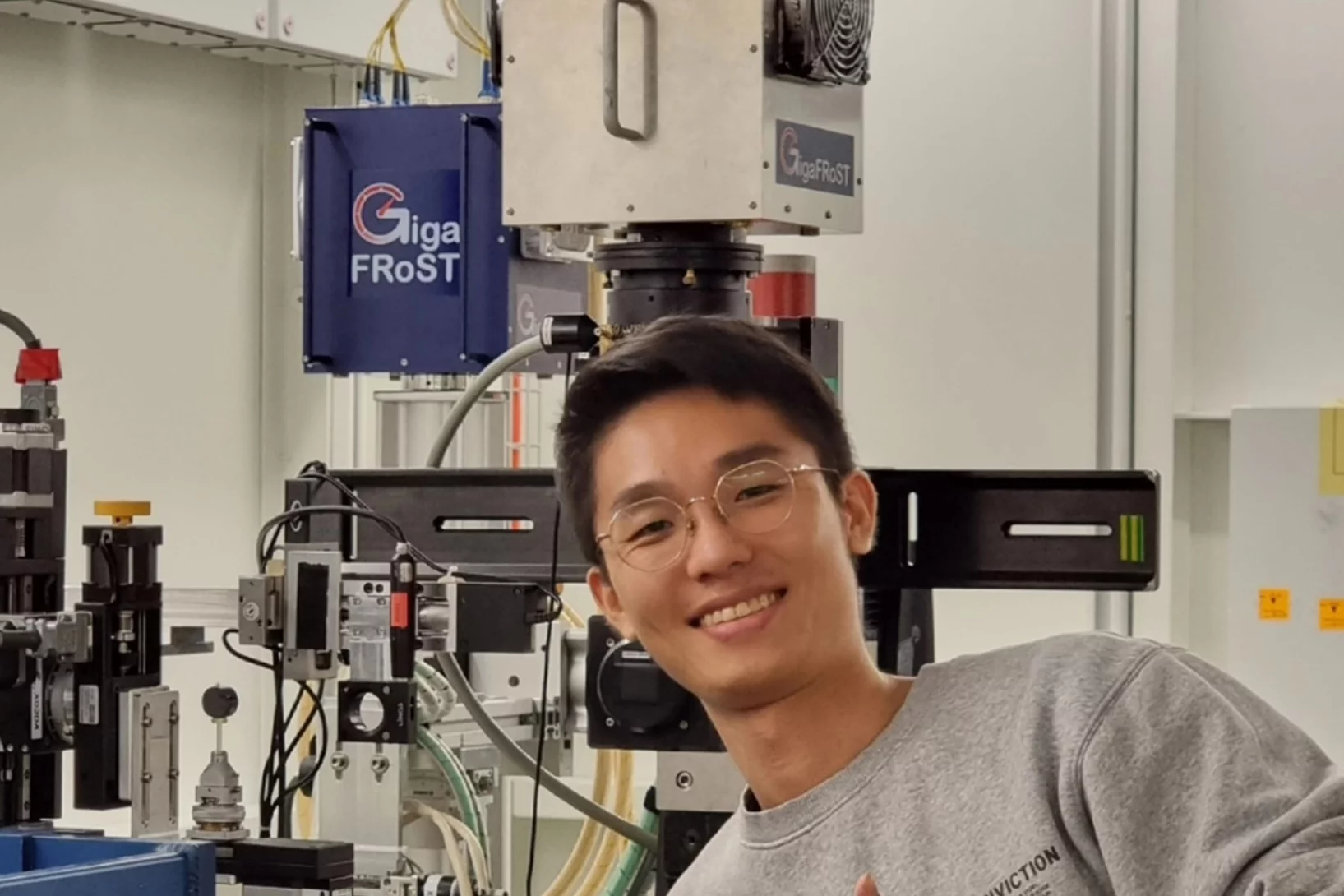
Jisoo Kim bags the 2022 Werner Meyer-Ilse Award
Jisoo Kim was awarded the 2022 Werner Meyer-Ilse Memorial Award. The WMI Award is given to young scientists for exceptional contributions to the advancement of X-ray microscopy through either outstanding technical developments or applications, as evidenced by their presentation at the International Conference on X-ray Microscopy and supporting publications. Jisoo was awarded for his development of the method "Time-resolved x-ray scattering tomography for rheological studies", and is co-recipient of the award with Yanqi Luo from the Advanced Photons Source for her work on applications. The award was presented during the 15th International Conference on X-ray Microscopy XRM2022 hosted by the National Synchrotron Radiation Research Center (NSRRC) in Hsinchu, Taiwan on 19 - 24 June, 2022.
Scientific Highlights

Coloration virtuelle de tissus en 3D
Des scientifiques de l’Institut Paul Scherrer PSI ont développé un système d’IA qui colore, à la manière de l’histologie classique, les images grises issues de la micro-CT. A l’avenir, cette méthode pourrait permettre d’analyser les tissus en 3D plutôt que sous forme de coupes minces microscopiques.

Un cœur volumineux et des sens aiguisés: les clés du foisonnement explosif des premiers poissons
La reconstruction par rayons X d’un fossile vieux de 400 millions d’années illumine un moment clé de notre passé évolutif le plus reculé.

Un fossile maître du camouflage aux rayons X
Le procédé d’imagerie du PSI aide à décoder la stratégie de chasse d’un prédateur préhistorique.
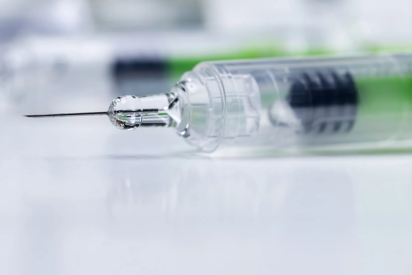
Du zinc détecté dans des seringues obstruées
Pour l'entreprise pharmaceutique MSD, ANAXAM a étudié, avec l'aide de scientifiques du PSI, si le zinc pouvait contribuer à l'obstruction des seringues préremplies.