Proton therapy has been used at PSI since 1984 to treat patients suffering from eye tumours. The therapy is carried out with the OPTIS radiation unit.
Preparation for Treatment
Before a definite decision is made for proton treatment, all patients are examined in detail at the referring eye clinic (Hôpital Ophtalmique Jules Gonin Lausanne , Eye clinic University of Essen , Eye clinic Innsbruck, Eye clinic University of Zürich). The referral of patients for proton therapy is fully in the competence of the ophthalmologist. Patients eligible for proton therapy will undergo surgery during which several small clips are sutured on the sclera (eye surface) in the proximity of the tumor. These clips will allow localization of tumor during the treatment with sub-millimeter accuracy. Patient arrives at PSI typically a few days after the surgery. Upon arrival, patient receives general information about treatment and undergoes steps necessary for the preparation of treatment. This includes creating an individual face mask with a small bite block, which helps the patient to keep his head completely still during treatment. In the next step, x-ray imaging of an eye is performed. These images are necessary for preparation of a treatment plan and for the later alignment of patient in front of the proton beam. After the imaging, patient leaves home and additional preparatory steps are performed in his absence. All prepared treatment plans are consulted with the referring ophthalmologists.
Administration of the Treatment
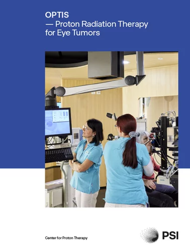
The treatment takes place at PSI on an out-patient basis, in the following week on 5 consecutive days and consists of a dry-run and a series of four treatments. During the dry-run, we verify whether there are not any limitations and patient positioning and treatment can be done in the fashion as intended in the treatment plan. The total dose delivered to the tumor is typically 60 Gy RBE. This dose is divided into four daily fractions of 15 Gy RBE. The irradiation itself lasts only about 60 seconds. However, the whole procedure requires about 20 minutes every day. Most of time is spend on the positioning of patient with the sub-millimeter accuracy. In order to keep the eye in the desired position for treatment, the patient must stare on an exactly calculated point of light. Before each treatment the position of the eye and the tumor are checked by means of x-ray images. The treatment is painless. In most cases the whole procedure, including surgery, takes approximately two to three weeks.

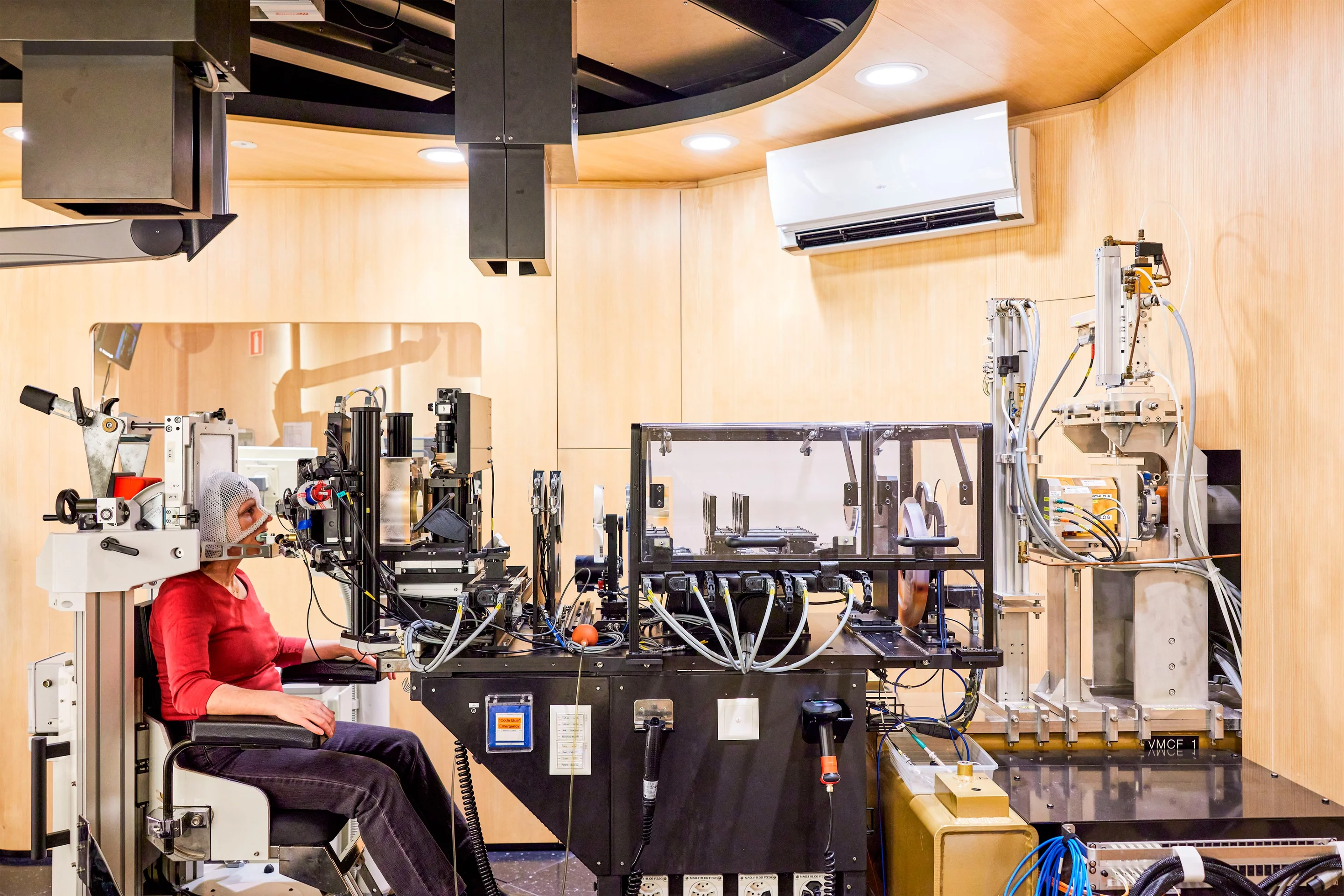
Central and indispensable: the collimator
When a patient enters the OPTIS treatment room for the first time, there is a lot to take in — and they might not even notice the shiny copper disc. Yet it plays a central role in eye irradiation: through the irregularly shaped hole in the middle, the proton beam emerges and targets the tumor in the eye.
The copper disc with the hole is called a collimator in technical language. The hole is milled so that its shape matches the contours of the tumor.

These apertures, as collimators are also known, are custom-made. They are milled in the PSI workshops and individually produced for each patient. The aperture, shaped according to the tumor, ensures that the proton beam only strikes the eye tumor and that the surrounding healthy tissue remains unaffected.