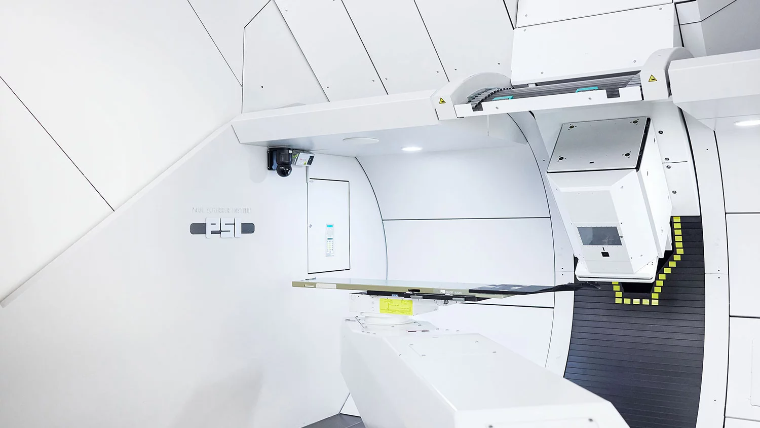
Gantry 2
At the new Gantry 2 the first patient was treated in late 2013. Technological innovations in this new treatment room are based on many years of experience gained in the development and operation of Gantry 1.
The speed of the beam delivery has been optimized : Gantry 2 uses two fast magnets with speeds of 1 to 2 cm / msec in the left-to-right and head-to-feet axes respectively to scan through the tumour. In the third dimension, the depth of penetration of the protons, the design of the beamline and gantry allows a change from one tumour layer to the next in about 100 msec (5 mm difference in proton range). Therefore “volumetric repainting” is possible, this means that during the same treatment the same volume is scanned through several times. This is an important approach to generate dose distributions that are less sensitive to organ motion.
The possibility of changing the beam intensity at high speed has also been implemented at the proton accelerator (cyclotron), although not yet used in patient treatments. This will enable so called "dose painting". Gantry 2 also has a parallel scanning system. However the size of the individual spots was reduced compared to Gantry 1 - even at low energies. The components in the nozzle are designed to have as little material as possible in the beam path. This maintains a small spot size at all energies (for 100-230 MeV, the width is <3-4 mm). The nozzle can be extended to reduce the air gap between the beam line exit window and the patient. The installation of collimators and compensators is also possible, if required.
A further notable development is the in-room imaging available: an in-room sliding CT is used for treatment planning and the daily verification of the patient position and includes the possibility to perform time-resolved imaging, "4D imaging". In addition, there is another x-ray system mounted on the gantry itself, which takes images in the direction of the proton beam ("beams eye view"). This provides increased precision and quality assurance, in particular in the treatment of moving tumours.