The Microspectroscopy Group has a long experience in synchrotron based hyperspectral imaging for a wide variety of materials and involving many different scientific disciplines. In particular we study the nanostructures of novel organic materials and the magnetic properties of thin films and multilayers employing a combination of X-ray microscopy and spectroscopy. We operate two beamlines at the SLS, PolLux and ISS. At these beamlines we perform our research and provide support for external users.
Recent Scientific Highlights and News:

Preserving film sound in the cold at the British Film Institute: new evidence for archives
Can magnetic film sound be stored in the cold? A collaboration between the British Film Institute and PSI found no detectable damage in the studied samples and points to a broader future for archives, in which laboratory methods and synchrotron techniques at the Swiss Light Source SLS help guide the preservation of complex audiovisual media.

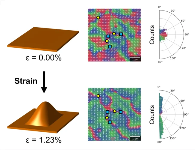
In-situ straining with a MEMS device
Researchers from an international collaboration between Switzerland, Italy, Germany, and Taiwan have developed a device for the in situ straining of freestanding ferroic films alllowing for ptychographic imaging whilst applying a mechanical strain. The results have been published as Editors' Suggestion within Physical Review B.
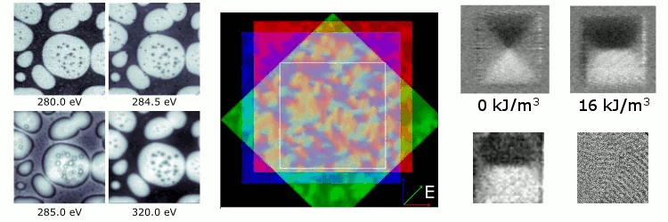
Designing antiferromagnetic domains by stretching membranes in STXM
Researchers from an international collaboration between the United Kingdom and Switzerland have performed imaging of an antiferromagnetic iron oxide membrane using soft X-ray microscopy. By stretching the membranes using a gas cell, the team investigated the modification of domain structures under strain.