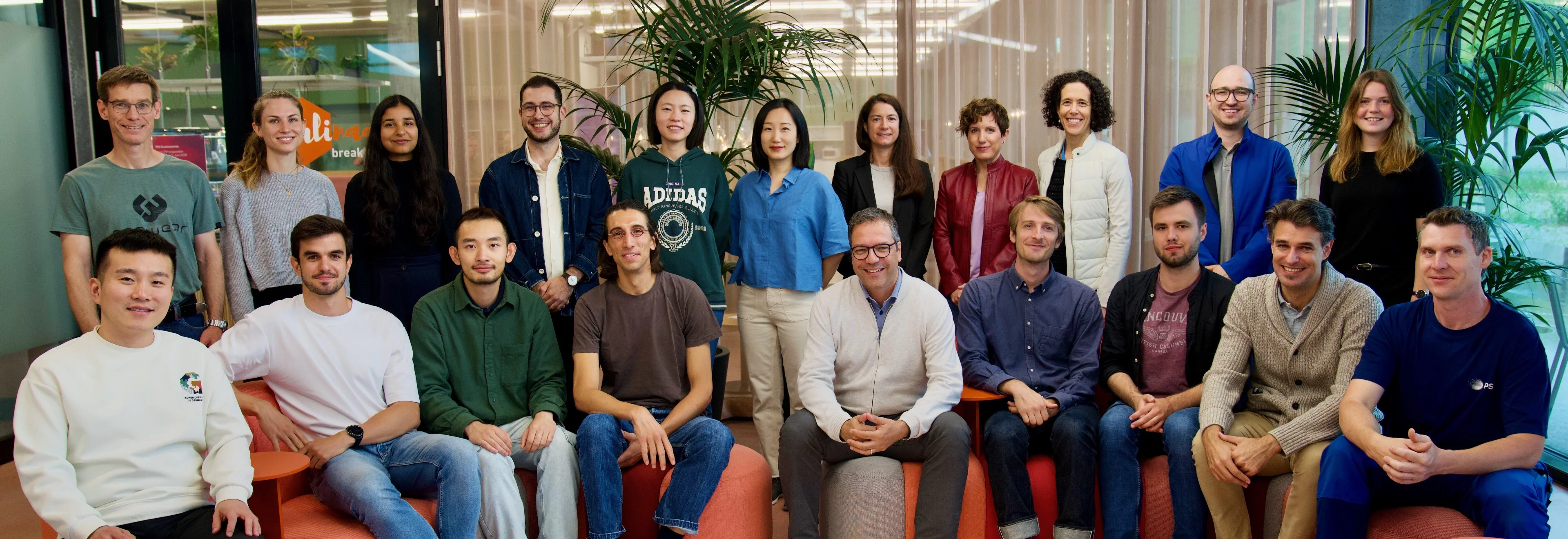
About us
Prof. Stampanoni heads a group of over 20 people, including four beamline scientists, one industrial liaison scientist, one technician, several scientists, many postdocs, PhD students, master students, and bachelor students. The team focuses on the development of tools, both instrumentation and algorithms, for tomographic X-ray imaging, exploiting synchrotron and laboratory sources. The group is engaged in the design and construction of ultra-fast data acquisition systems (stroboscopic coherent X-ray radiology and tomography) to provide dynamic investigation of rapidly evolving systems. The group also intensively develops optimized applications for fast, concurrent post-processing of tomographic data starting from simple normalization corrections to ad-hoc reconstruction and artifact reductions algorithms. Finally, the group investigates, creates and optimizes novel imaging modalities based on the coherent properties of synchrotron radiation and works on the translation of such work to conventional X-ray sources.
Associated Beamlines: TOMCAT 2.0 Multimodal Multiscale Dynamic Imaging
Scientific Highlights

Big heart, acute senses key to explosive radiation of early fishes
X-rays of a 400-million-year-old fossil illuminate a key moment in our deep evolutionary past.

X-rays reveal fossil stealth technology
PSI imaging helps to uncover the hunting strategy of a prehistoric predator.

Zinc detected in clogged syringes
With the help of researchers at PSI, ANAXAM has been investigating, on behalf of the pharmaceutical company MSD, whether zinc may contribute to clogging of pre-filled syringes.
Cause of clogged hypodermic needles discovered
Researchers at PSI and the ANAXAM technology transfer center have found the cause of clogging in prefilled syringes.
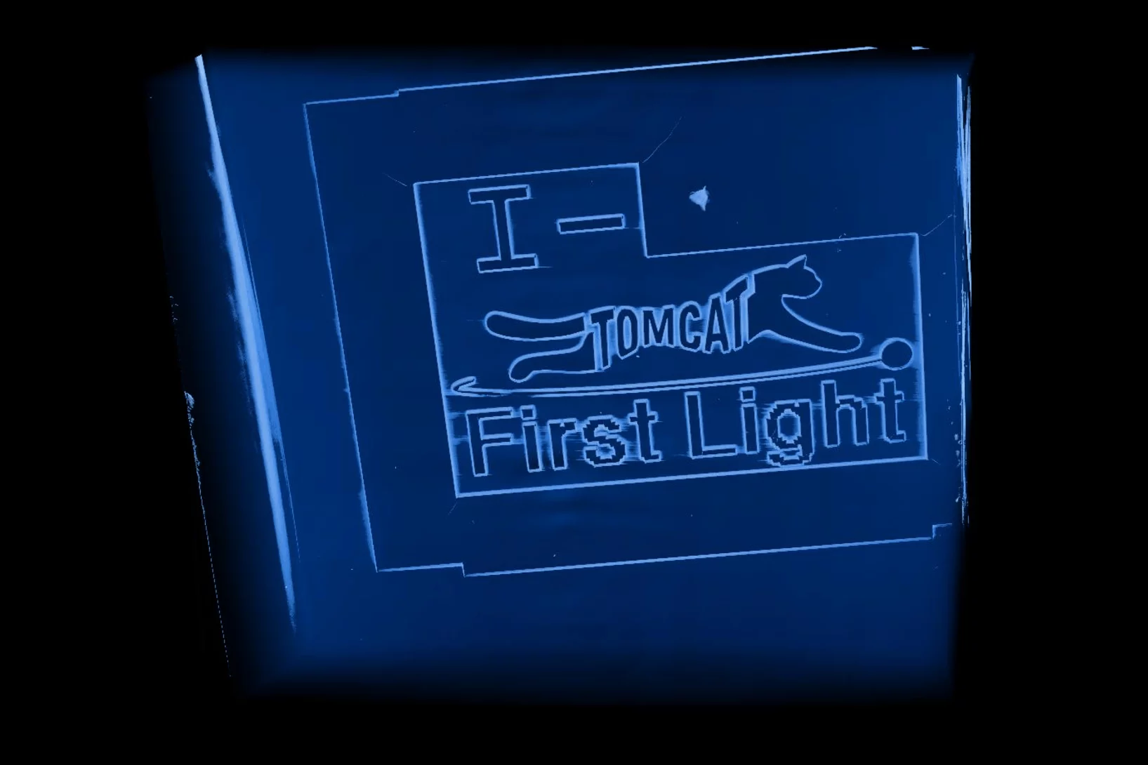
Brand new I-TOMCAT beamline comes to life
Marking another milestone in the TOMCAT 2.0 upgrade project, I-TOMCAT — our newly built beamline — received its first X-ray light on September 25, 2025.
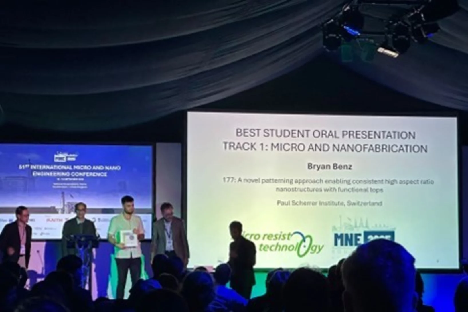
Bryan Benz awarded best talk at MNE 2025
Bryan Benz was awarded the conference’s best oral presentation at the 51st International Micro and Nano Engineering Conference (MNE 2025) for his talk entitled “A novel patterning approach enabling consistent high aspect ratio nanostructures with functional tops”.
S-TOMCAT beamline receives first X-ray light
On June 25th 2025, the S-TOMCAT beamline shutters were opened to receive first X-ray light from the new SLS 2.0 storage ring.
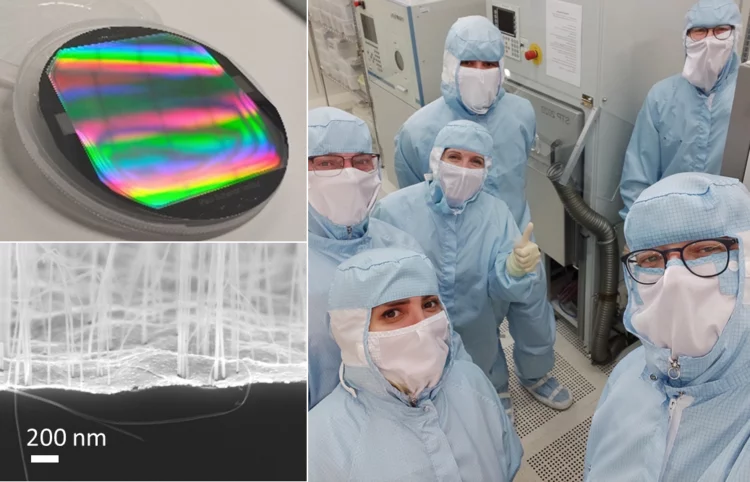
First vacuum-gas-controlled metal assisted chemical etching at 8-inch wafer scale
Our microfabrication team at Paul Scherrer Institut and the research institute Fraunhofer ENAS recently started a research cooperation in the field of gas-phase metal assisted chemical etching (MACE, MacEtch) technologies, supported by memsstar Ltd. Already in the early stages of the collaboration, the research team achieved very promising results with huge aspect ratio (at least 500:1) silicon nanowires, indicating an incredible development potential.