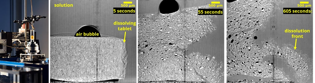
Reliable drug dissolution of immediate-release oral dosage forms is paramount for adequate in vivo performance and quality control. In this example, we have visualized the rapid dissolution of a Claritin tablet (Bayer Inc., Germany) by using time-resolved X-ray phase-contrast tomography at the TOMCAT beamline. A flow-through cell (see Figure 1a) was developed by ANAXAM to mimick flow conditions (2 mm/s) [1]. An indirect X-ray image detector composed of a 100 µm LuAG:Ce scintillator screen, a 4× magnification microscope (Optique Peter, France) and the GigaFRoST camera was used. The resulting pixel size of the image detector was 2.75 µm and the field of view 5.5 mm × 4.7 mm. The time evolution of tablet dissolution was recorded by acquiring a tomography scan every minute for 20 minutes. During this 20-minute duration the cell containing the sample rotates continuously at 0.33 rps (1 rotation per 3 seconds). Every tomography scan acquisition was acquired with 1500 projections as the cell completes 180 º rotation and takes 1.5 s. The exposure time per projection was 1 ms. This time resolution was chosen to be short enough to take a tomographic image of the dissolving tablet without motion blur. Figure 1b-1d shows the tomography slices at 5 seconds, 55 seconds and 605 seconds after the tablet goes in contact with an acidic solution. At first the tablet swelled, disintegrated, and then dissolved. X-ray phase-contrast tomography has allowed us to distinguish between different low density phases including water, air bubble, and individual components of the dissolving tablets.