
Show filters
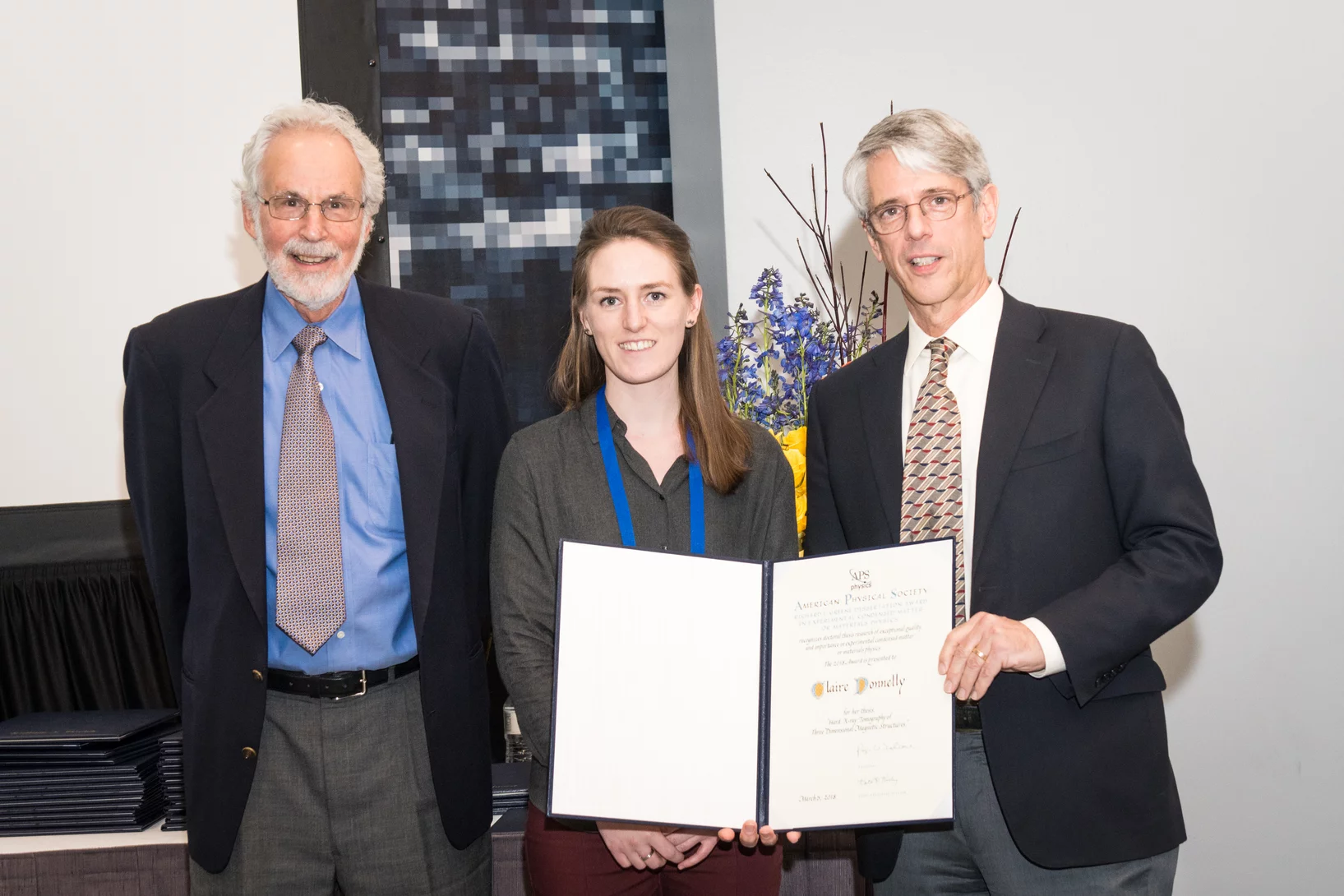
Claire Donnelly dissertation research awards
Claire Donnelly, Mesoscopic Systems (ETH Zurich - PSI), was awarded the COMSOL SPS Award in Computational Physics, the Werner Meyer-Ilse Memorial Award, the ETH Medal for an outstanding doctoral thesis, and the American Physical Society Richard L. Greene Dissertation Award.
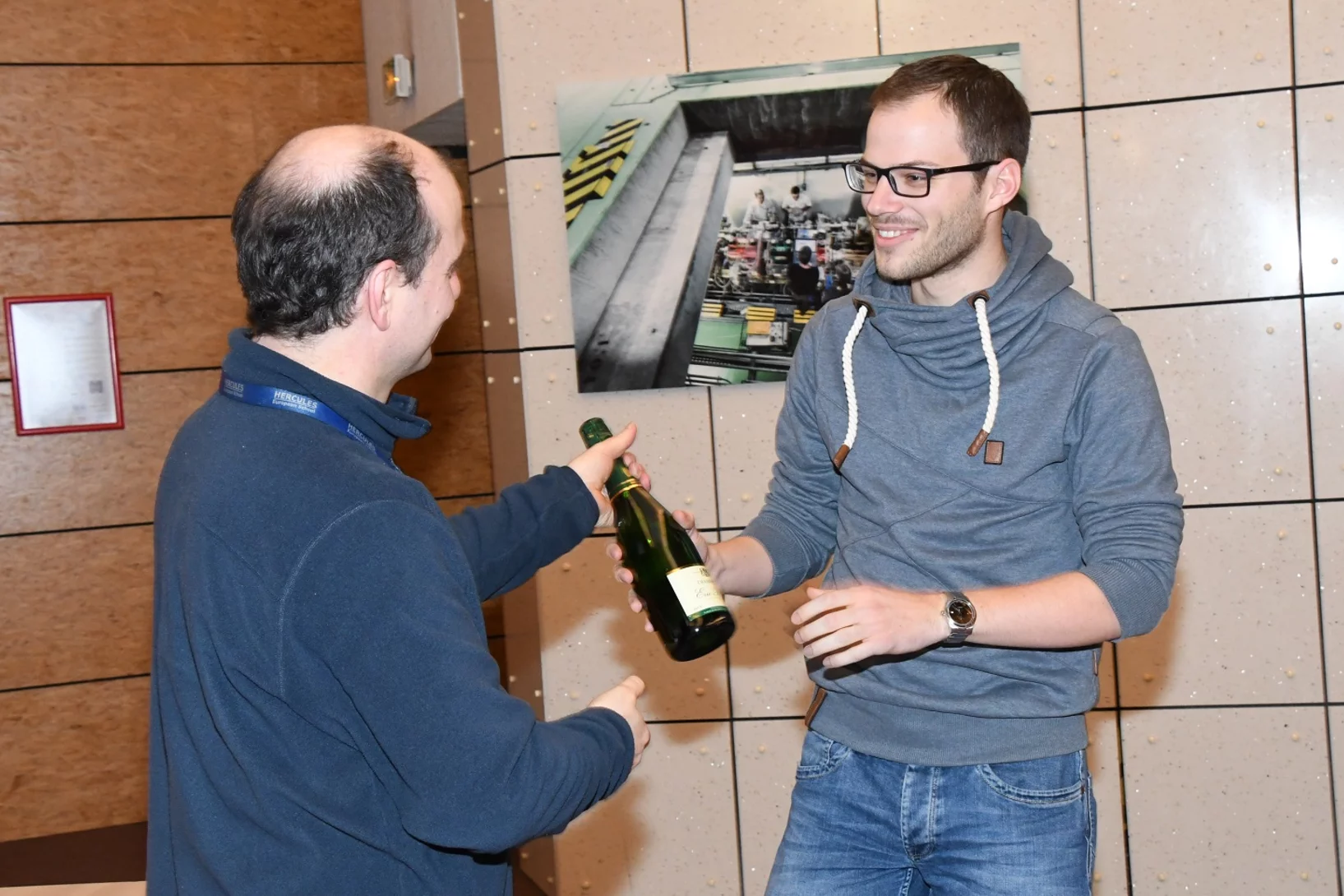
HERCULES School Poster Prize
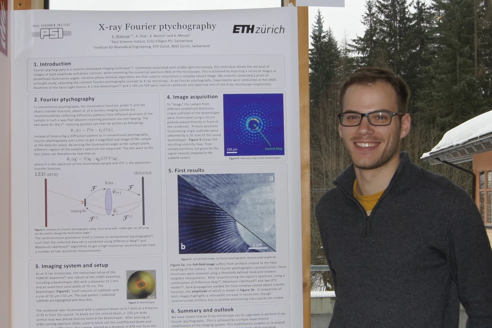
Klaus Wakonig was awarded the best poster prize in the 2018 rendition of the HERCULES European School in Grenoble, France. Klaus is currently a PhD student at the CXS group at PSI, developing X-ray Fourier ptychography.
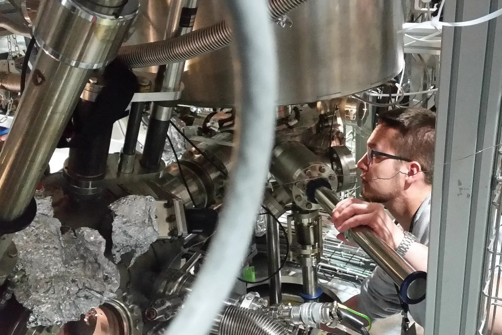
HERCULES at the Swiss Light Source
In the week of March 18-23 PSI welcomes 20 PhD students and postdocs taking part in the HERCULES 2018 school on Neutron and Synchrotron Radiation. They will attend lectures and perform two days of practical courses at several beam lines of the Swiss Light Source.
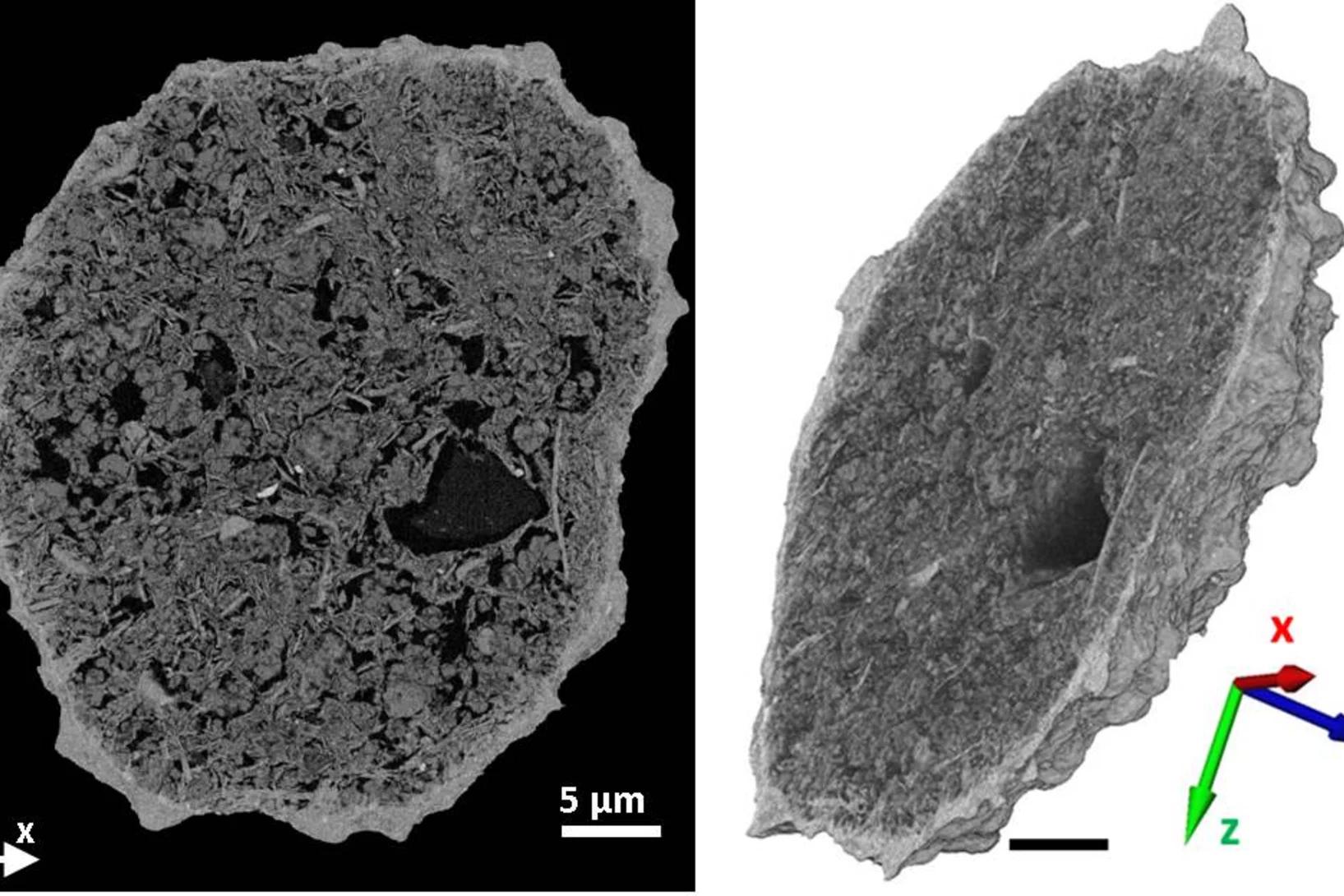
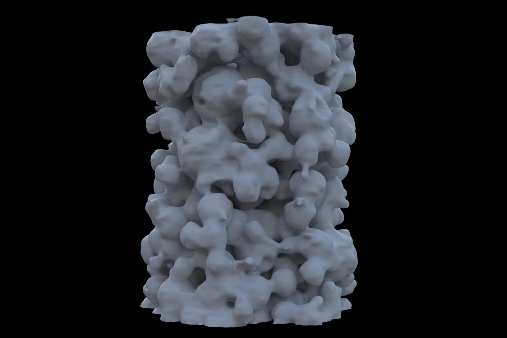
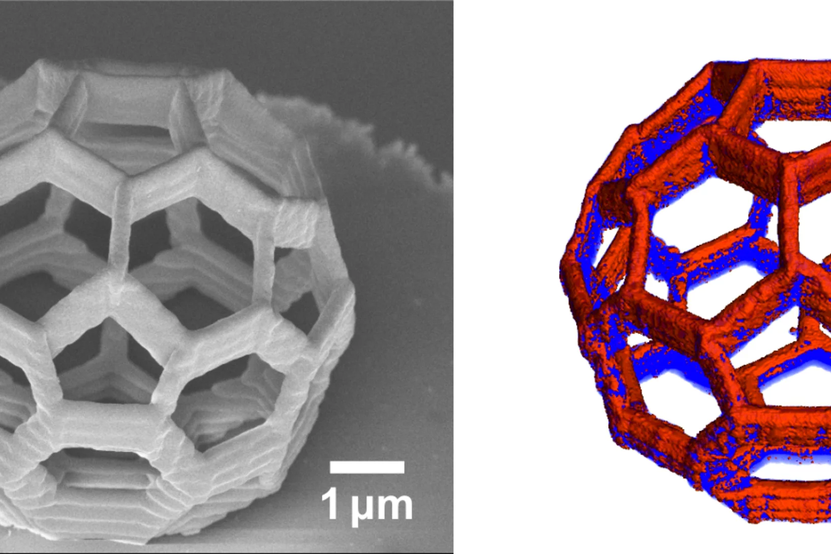
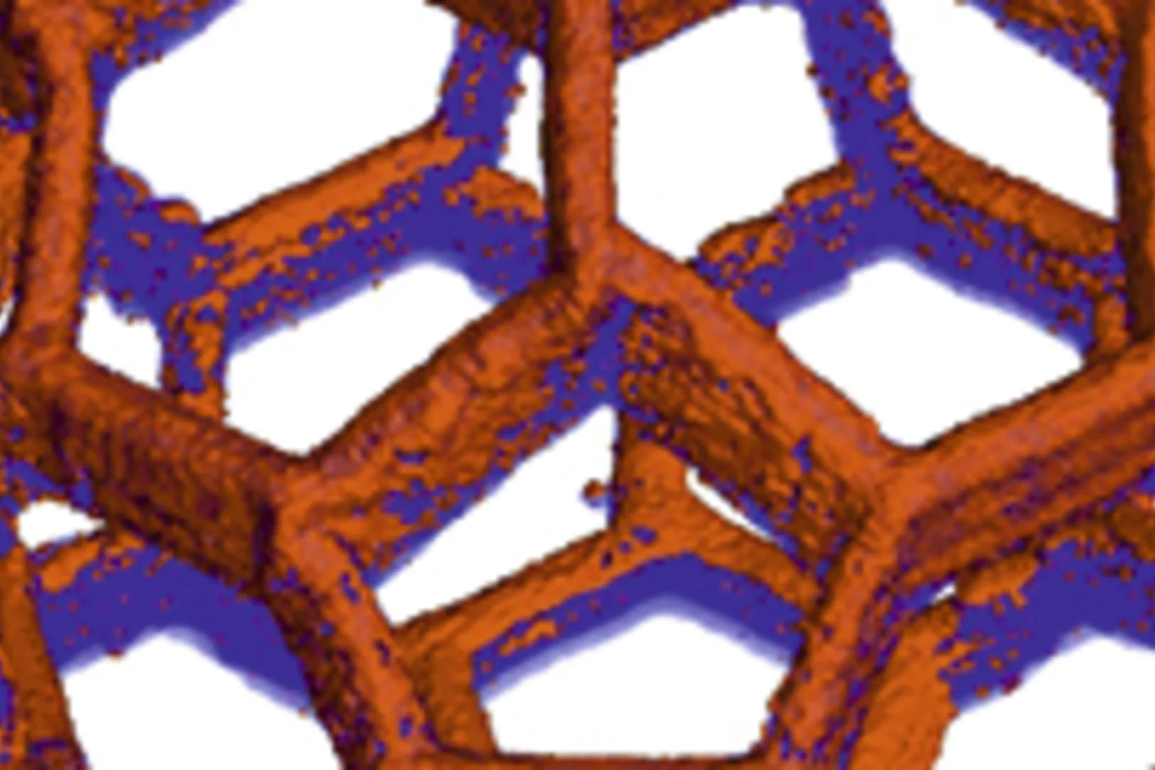
Making the world go round - a look into the structure of a prominent heterogeneous catalyst
Fluid catalytic cracking catalysts, which are composite particles of hierarchical porosity, were examined using ptychographic X-ray tomography. These particles are essential to the conversion of crude oil into gasoline. Examination of catalysts at decreasing levels of catalytic conversion efficacy allowed the detection of possible deactivation causes.
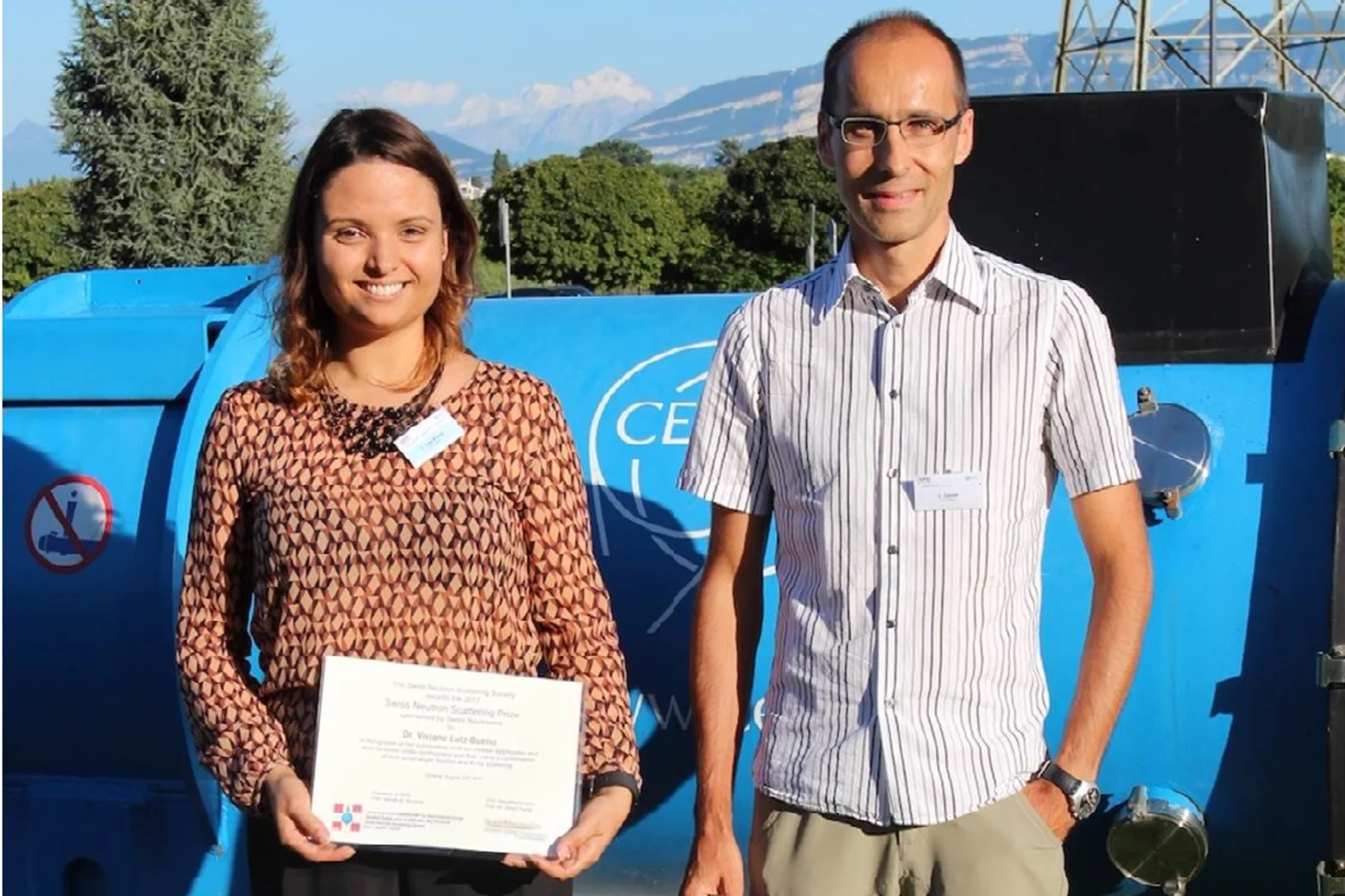
Swiss Neutron Scattering Prize
Viviane Lutz-Bueno was awarded the Swiss Neutron Scattering Prize of the Swiss Neutron Scattering Society, at the Joint Annual Meeting of the Swiss and Austrian Physical Society, for her PhD thesis Effects of formulation and flow on the structure of micellar aggregates carried out at ETH Zürich. Viviane is currently a postdoctoral fellow at the CXS group at PSI, developing scanning SAXS analysis methods.
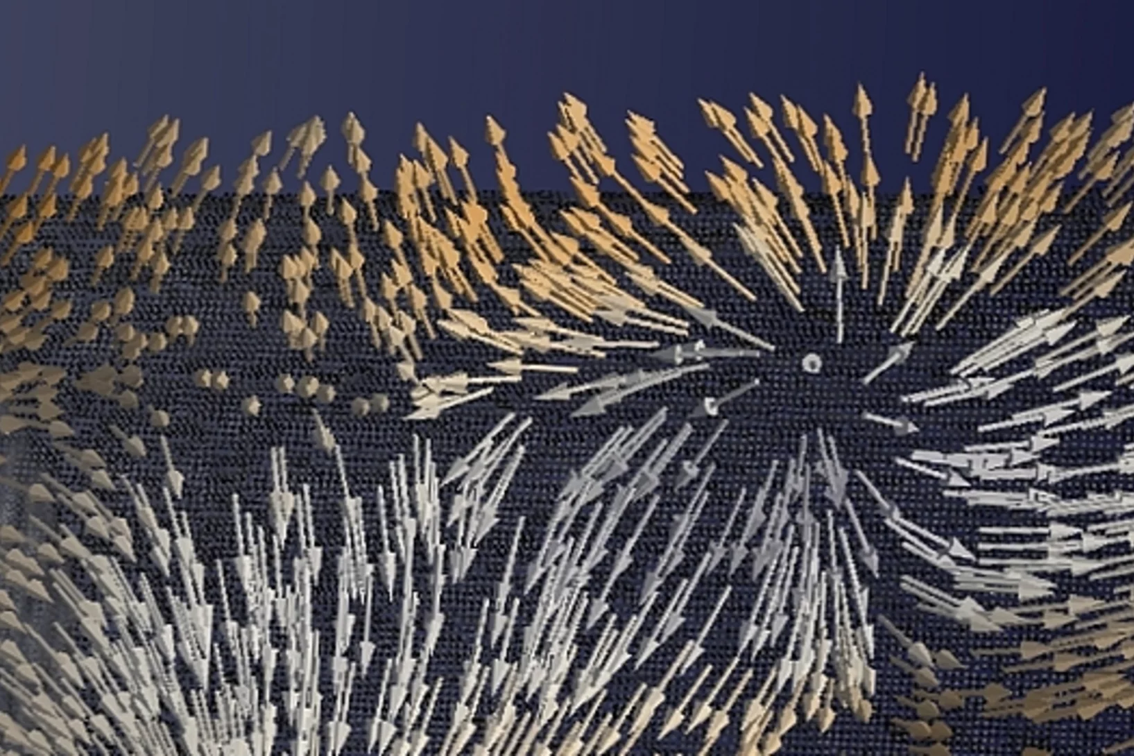
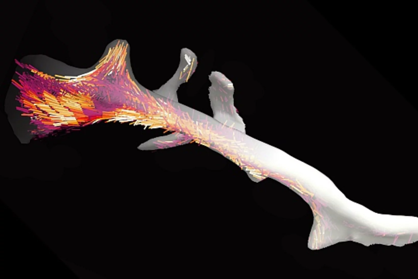
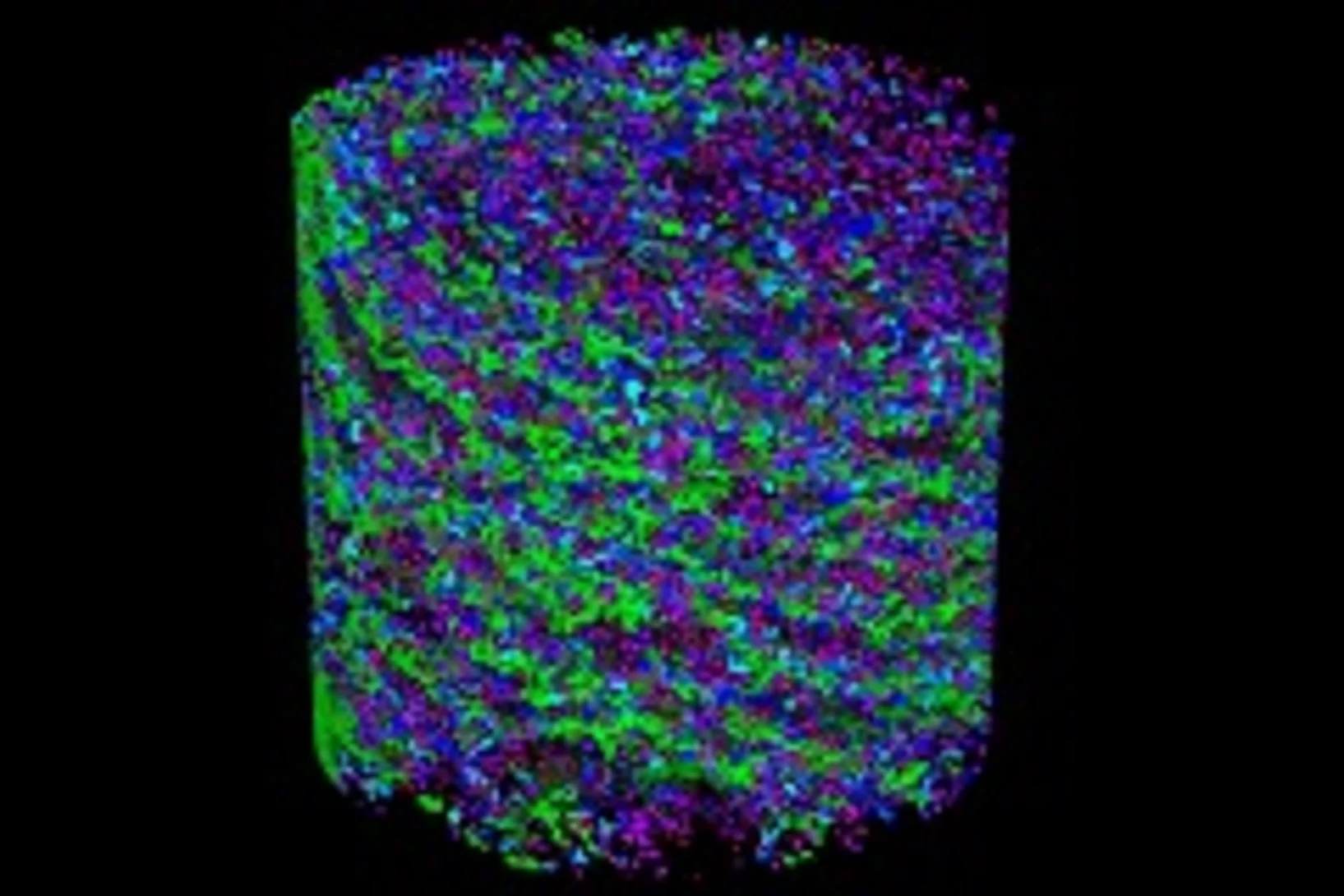
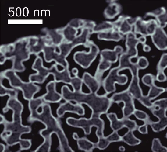
Diving into magnets
For the first time, scientists have made visible the directions of the magnetisation inside a 3D magnetic object. The smallest details in their visualisation were ten thousand times smaller than a millimetre. Among others, the magnetic structure contained one outstanding kind of pattern: magnetic singularities called Bloch points, which up to now were only known in theory.
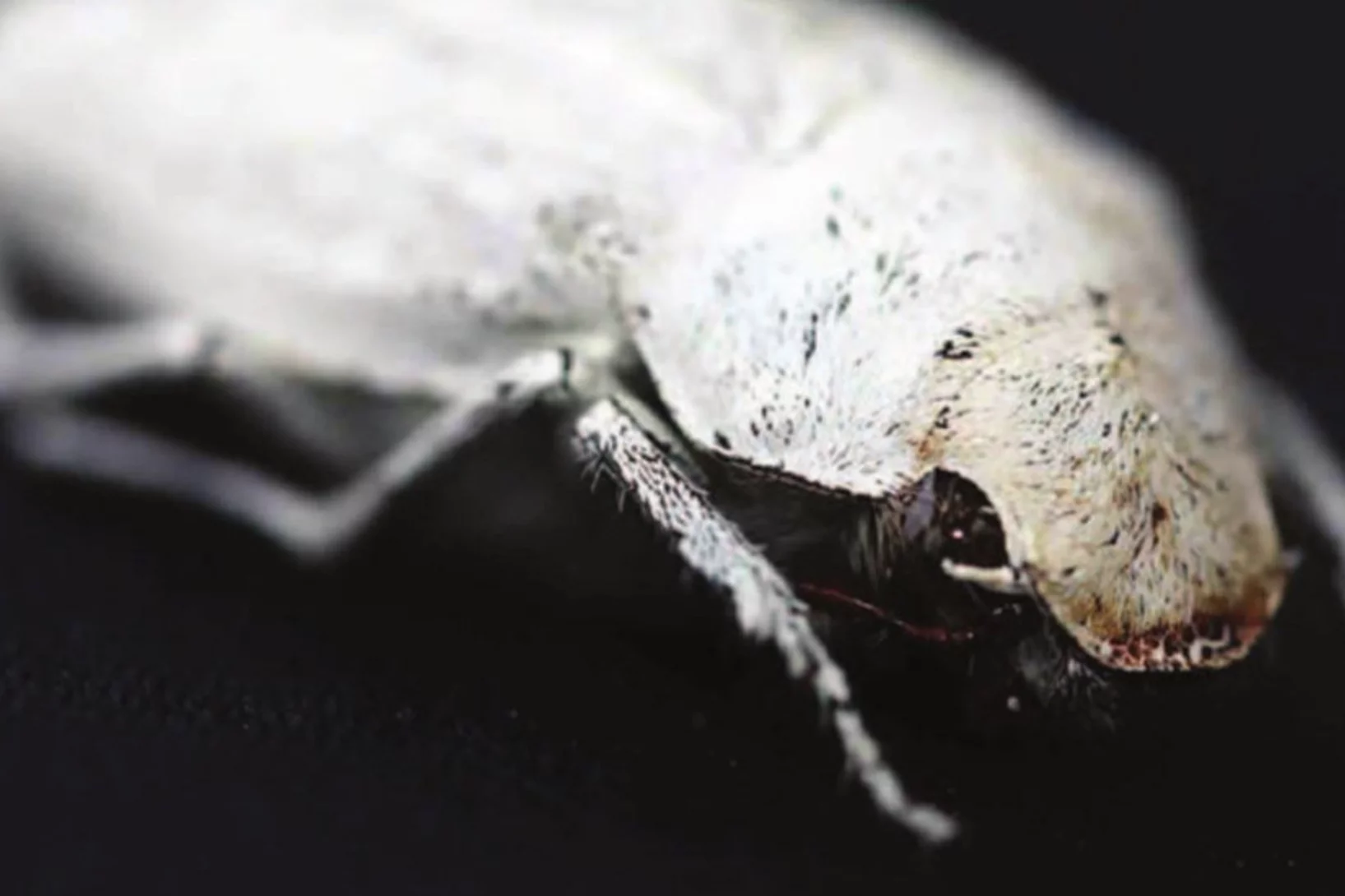
Photonic structure of white beetle wing scales: optimized by evolution
A very thin layer on this beetle’s wings exhibits a complicated structure on the nanoscale that gives them a bright white color. X-ray nanotomography acquired at the Swiss Light Source provides a faithful image of this structure in three dimensions with which scientists can confirm its evolutionary optimization: just enough material for an efficient reflection of white light.
European NESY Winterschool Young Scientist Best Poster Prize
Klaus Wakonig was awarded the "Young Scientist Best Poster Prize" along with a cash prize at the 10th European NESY Winterschool & Symposium on Neutron and Synchrotron Radiation. Klaus is a PhD student at the coherent X-ray scattering group (CXS) in PSI. His poster, entitled "X-ray Fourier ptychography," details his latest results in the implementation of Fourier ptychography at X-ray wavelengths for nanoimaging. Image credit ©NESY/Montanuniversitaet Leoben
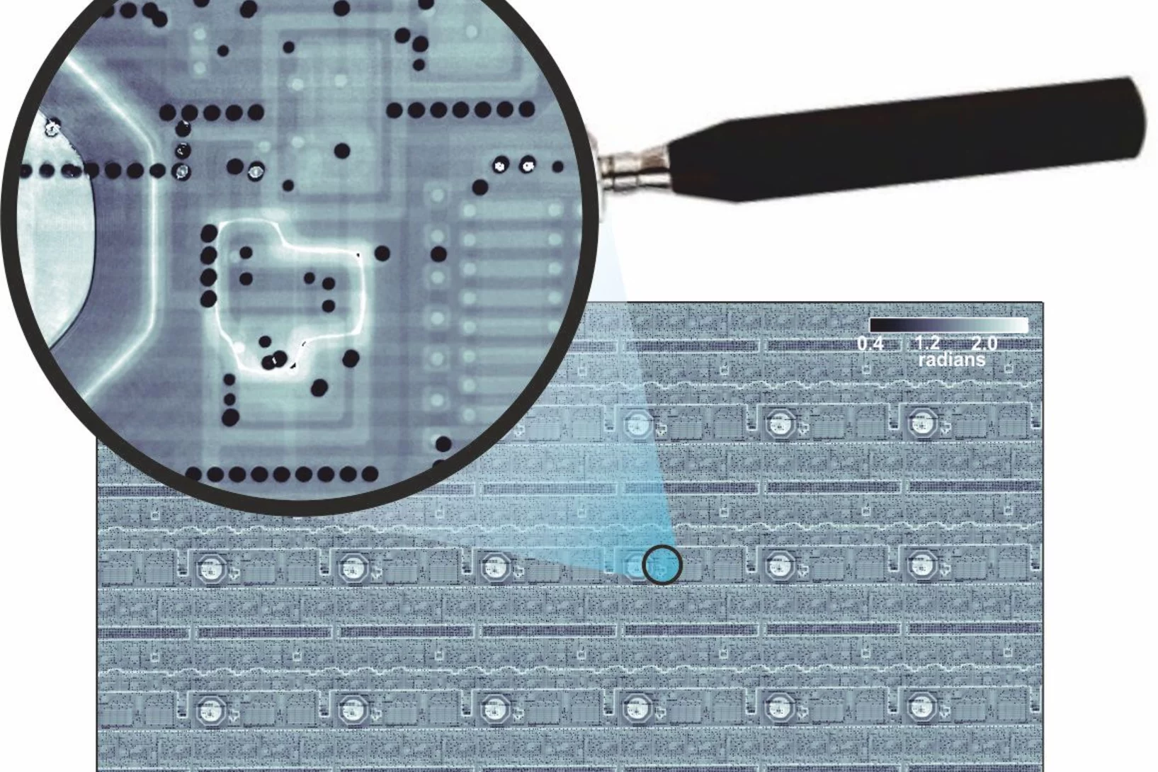
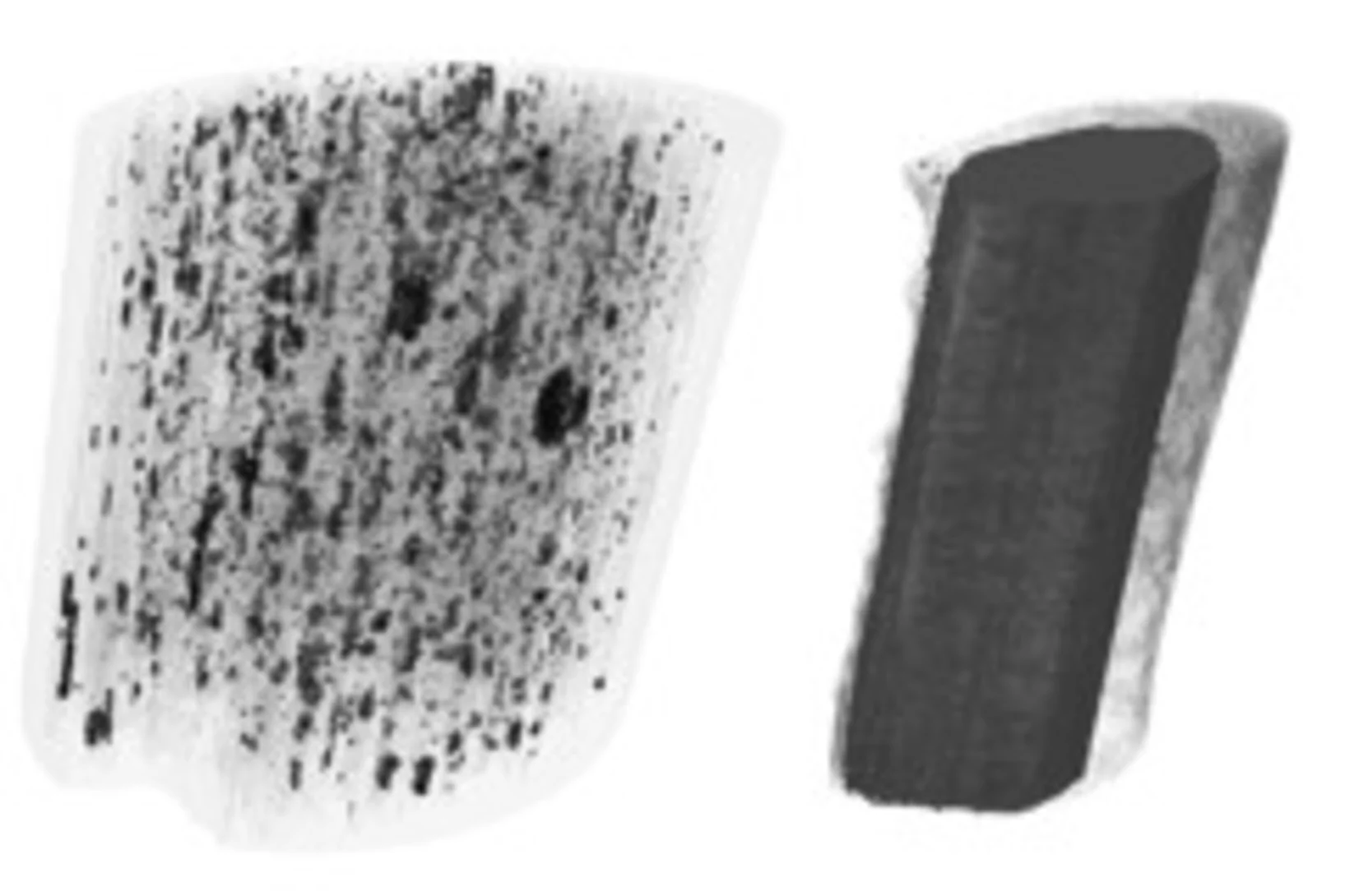
3-D X-ray imaging makes the finest details of a computer chip visible
Researchers at the PSI have made detailed 3-D X-ray images of a commercially available computer chip. In their experiment, they examined a small piece that they had cut out of the chip beforehand. This sample remained undamaged throughout the measurement. It is a major challenge for manufacturers to determine if, in the end, the structure of their chips conforms to the specifications. Thus these results represent one important application of an X-ray tomography method that the PSI researchers have been developing for several years.
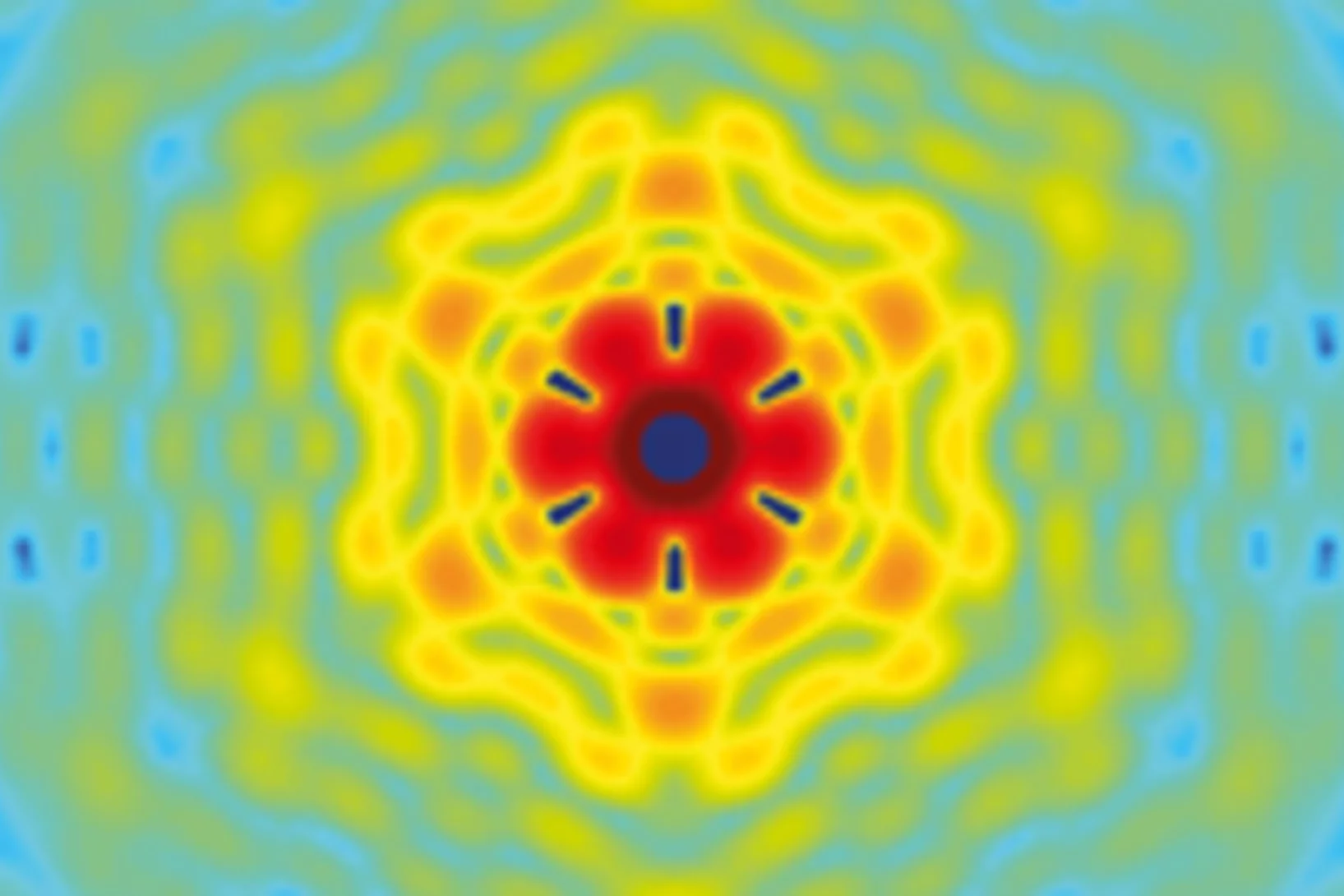
Interlaced zone plates push the resolution limit in x-ray microscopy
A novel type of diffractive lenses based on interlaced structures enable x-ray imaging at resolutions below 10 nm. The fabrication method and the test results of these novel x-ray lenses have been published in the journal Scientific Reports.
How does food look like on the nanoscale?
The answer to this question could save food industry a lot of money and reduce food waste caused by faulty production. Researchers from the University of Copenhagen and the Paul Scherrer Institut have obtained a 3D image of food on the nanoscale using ptychographic X-ray computed tomography. This work paves the way towards a more detailed knowledge of the structure of complex food systems.
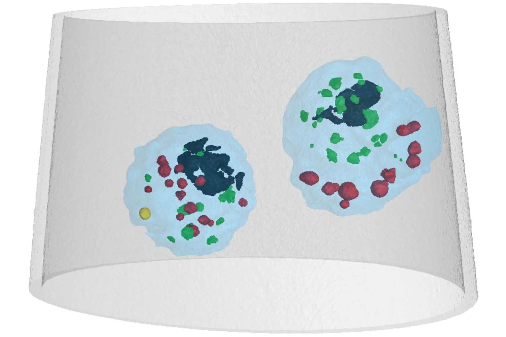
Mass density distribution of intact cell ultrastructure
The determination of the mass density of cellular compartments is one of the many analytical tools that biologists need to unravel the extremely complex structure of biological systems. Cryo X-ray nanotomography reveals absolute mass density maps of frozen hydrated cells in three dimensions.
3D nanostructure of a bone made visible
Bones are made up of tiny fibres that are roughly a thousand times finer than a human hair. Researchers at the Paul Scherrer Institute PSI have developed a new computer-based algorithm with which they were able to visualize the localised order and alignment of these nanostructures inside an entire piece of bone for the first time.
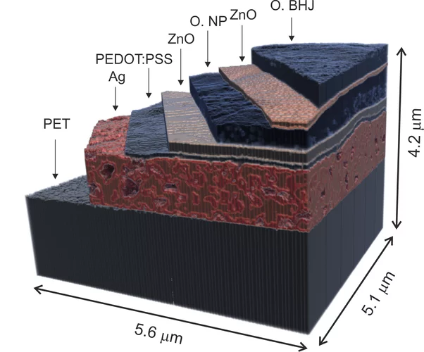
X-ray nanotomography aids the production of eco-friendly solar cells
Polymer solar cells are in the spotlight for sustainable energy production of the future. Characterization of these devices by X-ray nanotomography helps to improve their production using environmentally friendly materials.
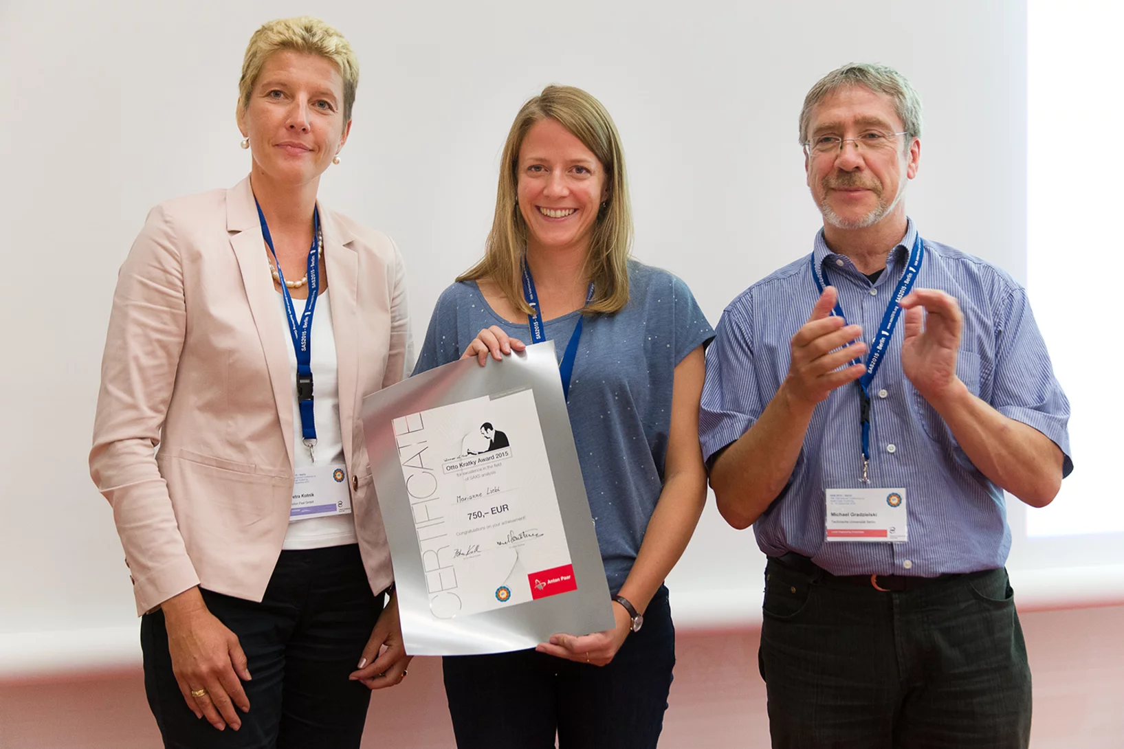
2015 Otto Kratky award
Marianne Liebi was awarded the 2015 Otto Kratky award by the Helmholtz-Centre Berlin for excellence in the field of small-angle X-ray scattering (SAXS) analysis. The award was bestowed in the last SAS2015 conference in Berlin. Marianne is a postdoctoral fellow in the coherent X-ray scattering group (CXS) in PSI, carrying out research in scanning SAXS measurement and analysis in 2D and 3D. Image credit ©HZB/Michael Setzpfandt
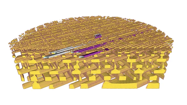
Element-Specific X-Ray Phase Tomography of 3D Structures at the Nanoscale
Recent advances in fabrication techniques to create mesoscopic 3D structures have led to significant developments in a variety of fields including biology, photonics, and magnetism. Further progress in these areas benefits from their full quantitative and structural characterization.
From inside an eggshell
Tiny cavities inside eggshells supply the materials that stimulate and control the shell’s growth. Using a novel imaging technique, researchers from the Paul Scherrer Institute (PSI), ETH Zurich and the Dutch FOM Institute AMOLF have succeeded in depicting these voids in 3D for the first time. In doing so, they lift an old limitation of tomographic images and hope that one day medicine will also benefit from their method.
Multiresolution X-ray tomography, getting a clear view of the interior
Researchers at PSI have developed a technique that combines tomography measurements at different resolution levels to allow quantitative interpretation for nanoscale tomography on an interior region of interest of the sample. In collaboration with researchers of the institute AMOLF in the Netherlands and ETH Zurich in Switzerland they showcase their technique by studying the porous structure within a section of an avian eggshell. The detailed measurements of the interior of the sample allowed the researchers to quantify the ordering and distribution of an intricate network of pores within the shell.
Nanometres in 3D
Scientists at the Paul Scherrer Institute and ETH Zurich have created 3D images of tiny objects showing details down to 25 nanometres. In addition to the shape, the scientists determined how particular chemical elements were distributed in their sample and whether these elements were in a chemical compound or in their pure state.

Innovation Award on Synchrotron Radiation 2014 for high-resolution 3D hard X-ray microscopy
The 2014 Innovation Award on Synchrotron Radiation was bestowed to researchers Ana Diaz, Manuel Guizar-Sicairos, Mirko Holler, and Jörg Raabe from the Paul Scherrer Institut, Switzerland, for their contributions to method and instrumentation development, which have set new standards in high-resolution 3D hard X-ray microscopy.
Fast scanning coherent X-ray imaging using Eiger
The smaller pixel size, high frame rate, and high dynamic range of next-generation photon counting pixel detectors expedites measurements based on coherent diffractive imaging (CDI). The latter comprises methods that exploit the coherence of X-ray synchrotron sources to replace imaging optics by reconstruction algorithms. Researchers from the Paul Scherrer Institut have recently demonstrated fast CDI image acquisition above 25,000 resolution elements per second using an in-house developed Eiger detector. This rate is state of the art for diffractive imaging and even on a par with the fastest scanning X-ray transmission instruments. High image throughput is of crucial importance for both materials and biological sciences for studies with representative population sampling.
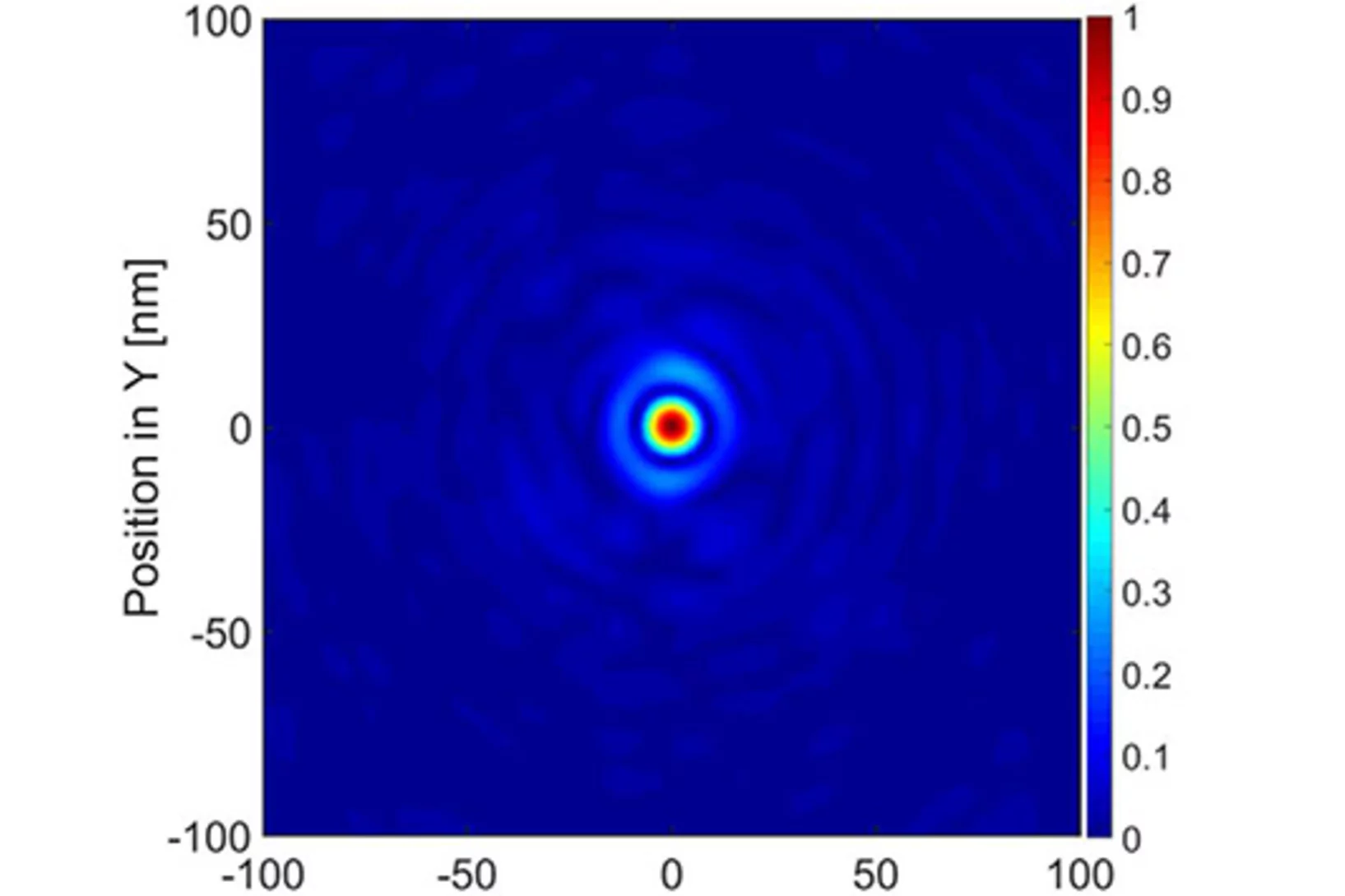
X-ray tomography reaches 16 nm isotropic 3D resolution
Researchers at PSI reported a demonstration of X-ray tomography with an unmatched isotropic 3D resolution of 16 nm in Scientific Reports. The measurement was performed at the cSAXS beamline at the Swiss Light Source using a prototype instrument of the OMNY (tOMography Nano crYo) project. Whereas this prototype measures at room temperature and atmospheric pressure, the OMNY system, to be commissioned later this year, will provide a cryogenic sample environment in ultra-high vacuum without compromising imaging capabilities. The researchers believe that such a combination of advanced imaging with state-of-the-art instrumentation is a promising path to fill the resolution gap between electron microscopy and X-ray imaging, also in case of radiation-sensitive materials such as polymer structures and biological systems.
Unique insight into carbon fibers on the nanoscale
The investigation of the mechanical properties of carbon fibers benefits from highly resolved three-dimensional density maps within representative volumes, but such images are not easily obtained with standard methods. Scientists from the Paul Scherrer Institut in collaboration with Honda R&D in Germany have recently visualized density distributions on the sub-micrometer scale within entire carbon fiber sections, revealing surprising graphite distributions within the fibers. This capability will prove useful for the systematic characterization of fibers, contributing to the development of light and robust materials at lower costs.
X-ray Laser: A novel tool for structural studies of nano-particles
Prominent among the planned applications of X-ray free electron laser facilities, such as the future SwissFEL at the Paul Scherrer Institute, PSI, are structural studies of complex nano-particles, down to the scale of individual bio-molecules. A major challenge for such investigations is the mathematical reconstruction of the particle form from the measured scattering data. Researchers at PSI have now demonstrated an optimized mathematical procedure for treating such data, which yields a dramatically improved single-particle structural resolution. The procedure was successfully tested at the Swiss Light Source synchrotron at PSI.
Imaging fluctuations with X-ray microscopy
X-rays are used to investigate nanoscale structures of objects as varied as single cells or magnetic storage media. Yet, high-resolution images impose extreme constraints on both the X ray microscope and the samples under investigation. Researchers at the Technische Universität München the PSI now showed how to relax these conditions without loss of image quality. They further showed how to image objects featuring fast fluctuations, such as the rapid switching events that determine the life time of data storage in magnetic materials.
Nanoforscher untersuchen Karies
Forscher der Universität Basel und des Paul Scherrer Instituts konnten im Nanomassstab zeigen, wie sich Karies auf die menschlichen Zähne auswirkt. Ihre Studie eröffnet neue Perspektiven für die Behandlung von Zahnschäden, bei denen heute nur der Griff zum Bohrer bleibt. Die Forschungsergebnisse wurden in der Fachzeitschrift «Nanomedicine» veröffentlicht.This news release is only available in German.
X-ray methods help to understand brain disorders better
An international team of researchers has developed a new method for making detailed X-ray images of brain tissue, which has been used to make the myelin sheaths of nerve fibres visible. Damage to these protective sheaths can lead to various disorders, such as multiple sclerosis. The facility for creating these images of the protective sheaths of nerve cells is being operated at the Swiss Light Source (SLS), at the Paul Scherrer Institute.
High-resolution method for computed nano-tomography developed
High-resolution method for computed nano-tomography developedA novel nano-tomography method developed by a team of researchers from the Technische Universität München, the Paul Scherrer Institute and the ETH Zurich opens the door to computed tomography examinations of minute structures at nanometer resolutions. The new method makes possible, for example, three-dimensional internal imaging of fragile bone structures.