• Paul Scherrer Institut PSI
  • PSI Research, Labs & User Services

Digital User Office

  • Digital User Office
  • DE
  • EN
  • FR
Paul Scherrer Institut (PSI)
Search
Paul Scherrer Institut (PSI)

Hauptnavigation

  • Research at PSIOpen mainmenu item
    • Research Initiatives
    • Ethics and Research integrity
    • Scientific Highlights
    • Scientific Events
    • Scientific Career
    • PSI-FELLOW
    • PSI Data Policy
  • Research Divisions and LabsOpen mainmenu item
    • Overview
    • Research with Neutrons and Muons
    • Photon Science
    • Energy and Environment
    • Nuclear Energy and Safety
    • Biology and Chemistry
    • Scientific Computing, Theory and Data
    • Large Research Facilities
  • Facilities and InstrumentsOpen mainmenu item
    • Overview
    • Large Research Facilities
    • Facilities
    • PSI Facility Newsletter
  • PSI User ServicesOpen mainmenu item
    • User Office
    • Methods at the PSI User Facilities
    • Proposals for beam time
    • Proposal Deadlines
    • Data Analysis Service (PSD)
    • EU support programmes
  • New ProjectsOpen mainmenu item
    • SLS 2.0
    • IMPACT
  • DE
  • EN
  • FR

Digital User Office (mobile)

  • Digital User Office

You are here:

  1. PSI Home
  2. Labs & User Services
  3. PSD
  4. LSB
  5. CXS
  6. Photonic structure of white beetle wing scales: optimized by evolution

Secondary navigation

Coherent X-Ray Scattering Group

  • People
  • Research
  • Scientific Highlights and News
  • Publications
22 June 2017

Photonic structure of white beetle wing scales: optimized by evolution

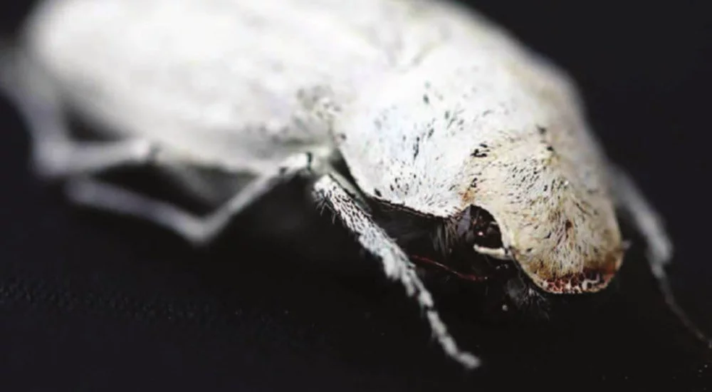
Figure 1: Cyphochilus white beetle

Cyphochilus white beetles (see Fig. 1) have developed a complicated three-dimensional (3D) photonic structure on their wing scales in order to efficiently reflect white light. At the same time, this structure is very porous and is confined within a thin layer of about 10  µm, about one fifth of the thickness of ordinary white paper, which makes it very light and therefore advantageous to fly. Researchers of the University of Fribourg and their collaborators wanted to understand how this fascinating structure is optimized, for which they needed a faithful 3D image. However, conventional microscopy techniques providing enough spatial resolution such as electron microscopy required the sample to be cut for imaging consecutive slices, causing damage of the structure during the process.

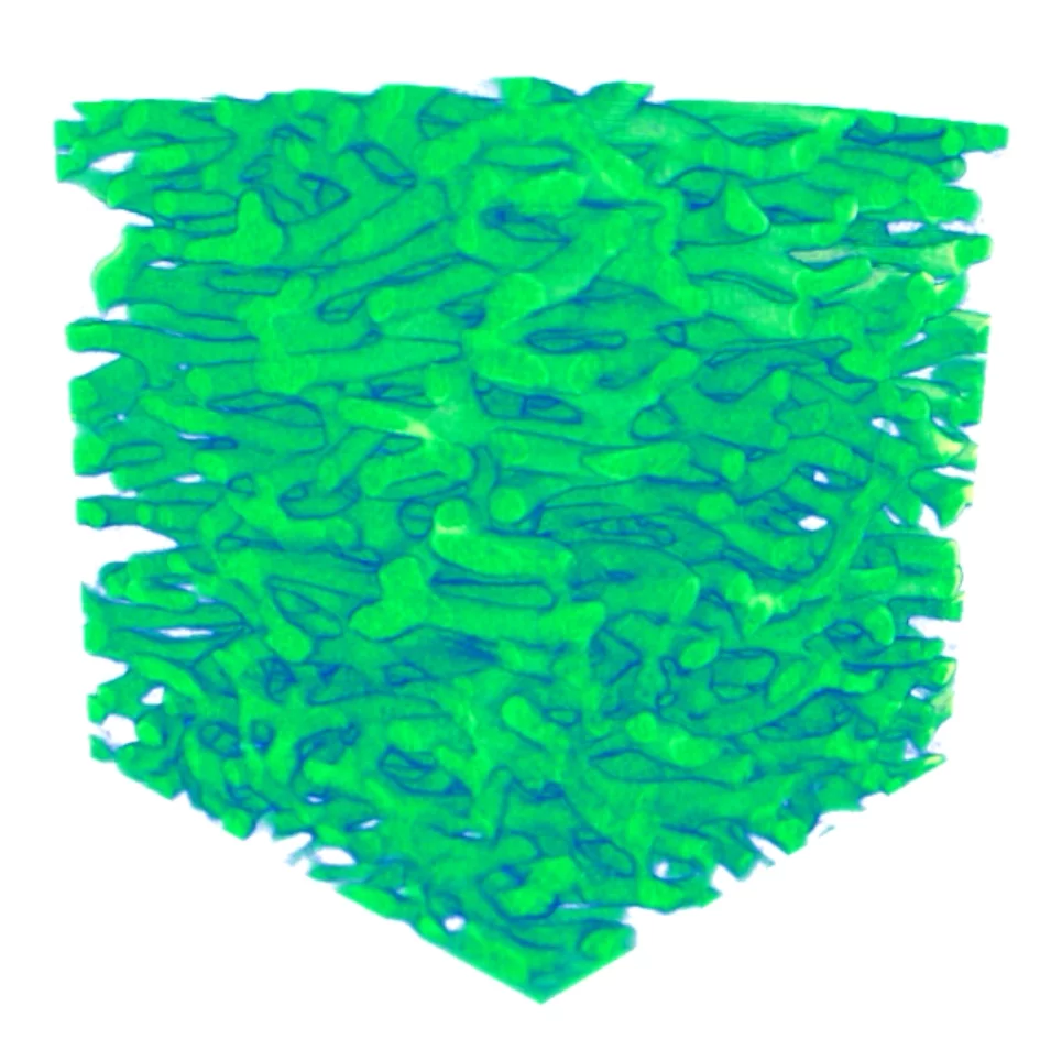
Figure 2: 3D rendering of chitinous fiber network of about 7×7×7 µm3 size

The tOMography Nano crYo (OMNY) imaging system operated at the coherent small-angle X-ray scattering (cSAXS) beamline at the Swiss Light Source was the key instrument that allowed them to image a small volume of the sample for their investigations. This equipment can keep the specimen at a temperature of 90 K to minimize structural changes caused by radiation damage while scanning the sample with respect to the beam with a precision of about 10 nm, which was needed to acquire a 3D image of 28 nm resolution, as shown in Fig. 2, by ptychographic X-ray computed tomography [1]. This is achieved by an ingenious system based on differential optical interferometry compatible with sample rotation which had been already tested for radiation hard samples at room temperature [2].

Optical reflectivity simulations directly performed on the measured structure confirmed that the structure developed by this beetle was indeed optimized for an efficient reflection of white light while using as little material as possible. Indeed, increasing the size or thickness of the struts in the structure or stretching the entire structure did not improve the reflection efficiency significantly, while the opposite was clearly detrimental. Humans could now take advantage of this evolutionary-optimized structure for applications e.g. in the food industry.

References
[1] M. Dierolf et al., Nature 467, 436 (2010) DOI: 10.1038/nature09419
[2] M. Holler et al., Sci. Rep. 4, 3857 (2014) DOI: 10.1038/srep03857
Additional information
Taking Color Inspiration from Evolutionary-Optimized White Beetle Wings, Advanced Science News
Contact
Dr. Ana Diaz
Beamline Scientist, Swiss Light Source
Paul Scherrer Institut
Telephone: +41 56 310 5626
E-mail: ana.diaz@psi.ch
Original Publication
Evolutionary-Optimized Photonic Network Structure in White Beetle Wing Scales
B. D. Wilts, X. Sheng, M. Holler, A. Diaz, M. Guizar-Sicairos, J. Raabe, R. Hoppe, S.-H. Liu, R. Langford, O. D. Onelli, D. Chen, S. Torquato, U. Steiner, C. G. Schroer, S. Vignolini, A. Sepe
Adv. Mater. 30, 1702057 (2018)
DOI: 10.1002/adma.201702057
Sidebar

Associated Beamlines

  • cSAXS

Contact

Dr. Andreas Menzel
Paul Scherrer Institut
5232 Villigen-PSI
Switzerland
Telephone: +41 56 310 3711
E-mail: andreas.menzel@psi.ch

Photon Science Division

Homepage of PSI Division Photon Science (PSD)


top

Footer

Paul Scherrer Institut

Forschungsstrasse 111
5232 Villigen PSI
Switzerland

Telephone: +41 56 310 21 11
Telefax: +41 56 310 21 99

How to find us
Contact

Visitor Centre psi forum
School Lab iLab (in German)
Center for Proton Therapy
PSI Education Centre
PSI Guest House
PSI Gastronomie (in German)
psi forum shop

Service & Support

  • Phone Book
  • User Office
  • Accelerator Status
  • PSI Publications
  • Suppliers
  • E-Billing
  • Computing
  • Safety (in German)

Career

  • Working at PSI
  • Job Opportunities
  • Training and further education
  • Career Center
  • Vocational Training (in German)
  • PSI Education Center

For the media

  • PSI in brief
  • Facts and Figures
  • Media corner
  • Media Releases
  • Social Media

Follow us: Twitter (in English) LinkedIn Youtube Facebook Instagram Issuu RSS

Footer legal

  • Imprint
  • Terms and Conditions
  • Editors' login