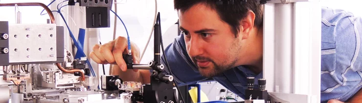
The Coherent X-ray Scattering (CXS) group develops techniques in scanning- and time-resolved SAXS and high-resolution scanning X-ray microscopy at the cSAXS beamline. In collaboration with research groups, within PSI and international universities and research institutes, we apply these techniques to a wide range of problems in the fields of biology, biomedical research and materials science.
We will open position at cSAXS for small-angle scattering tensor tomography in combination with ptychographic tomography. Contact us for details.
Scientific Highlights
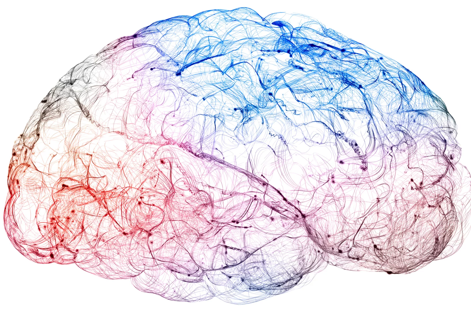
X-rays bring high-resolution brain mapping within reach
A new imaging breakthrough could reveal brain connectivity in 3D detail never before accessible.
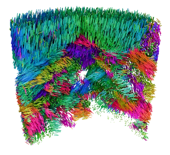
Mapping the Nanoscale Architecture of Functional Materials
A new X-ray technique reveals the 3D orientation of ordered material structures at the nanoscale, allowing new insights into material functionality.

New X-ray world record: Looking inside a microchip with 4 nanometre precision
Researchers at PSI have succeeded in imaging the spatial structure of a computer chip with a record resolution of 4 nanometres using X-rays.