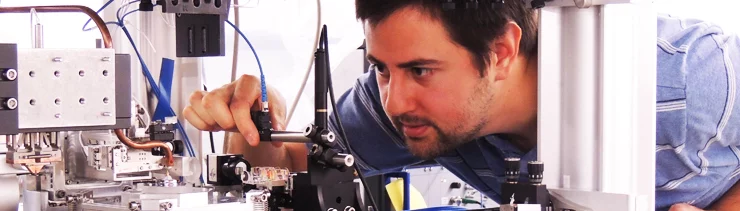
The Coherent X-ray Scattering (CXS) group develops techniques in scanning- and time-resolved SAXS and high-resolution scanning X-ray microscopy at the cSAXS beamline. In collaboration with research groups, within PSI and international universities and research institutes, we apply these techniques to a wide range of problems in the fields of biology, biomedical research and materials science.
We will open position at cSAXS for small-angle scattering tensor tomography in combination with ptychographic tomography. Contact us for details.
Scientific Highlights
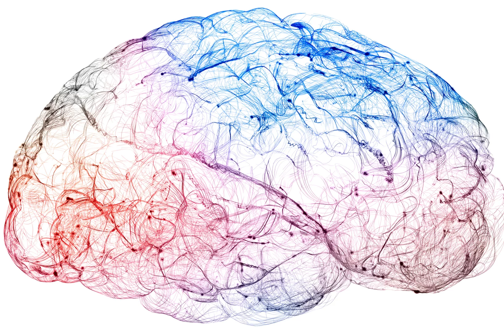
Hochauflösende Gehirnkartierung dank Röntgenlicht in Reichweite
Ein Durchbruch bei einem bildgebenden Verfahren könnte die Verbindungen innerhalb des Gehirns in bisher unerreichter 3D-Auflösung sichtbar machen.
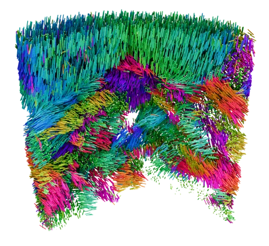
Mapping the Nanoscale Architecture of Functional Materials
A new X-ray technique reveals the 3D orientation of ordered material structures at the nanoscale, allowing new insights into material functionality.

Neuer Röntgenweltrekord: Blick in einen Computerchip auf 4 Nanometer genau
Mit einer Rekordauflösung von 4 Nanometern gelang es Forschenden am PSI, die räumliche Struktur eines Computerchips mithilfe von Röntgenlicht abzubilden.