Show filters
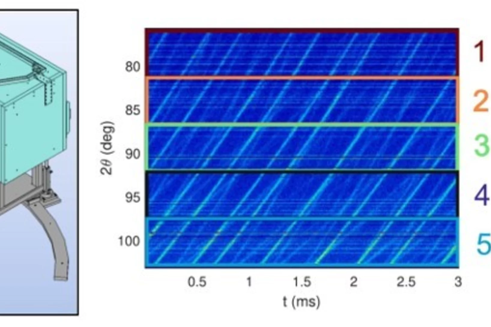
Texture analysis implementation at the neutron strain diffractometer POLDI
This study presents the implementation of a novel data analysis methodology to perform spatially resolved crystallographic texture analyses in bulk specimens at POLDI, the pulsed frame overlap diffractometer at SINQ, Paul Scherrer Institute. The method is based on the determination of several incomplete pole figures. To increase the angular resolution, the POLDI diffraction bank is split into several virtual units of smaller angular coverage. The diffraction data of each virtual unit can then be analyzed individually and used to create experimental pole figures from the Euler angles of the explored sample orientations. Additionally, to help the analyses, a new numerical tool was developed and implemented at POLDI to calculate neutron flight path of each virtual detector as a function of sample size, geometry, and orientation. Leveraging on the SALOME platform’s Geom module (open-source CAD modeler), the tool allows inserting CAD objects into a virtual detailed PODI geometry. This allows to automate sample positioning and orientation within the instrument frame and computes flight path intersections. It serves two main purposes: enhancing texture analysis through precise path calculations and aiding experimental design by visually evaluating orientation feasibility and estimating counting times. Finally, to complete the analysis path from the experiments to the results, the experimental and numerical evaluations are processed together with POLTex (MATLAB-based toolbox) to obtain the orientation distribution functions. To demonstrate the analysis routine, the crystallographic texture of an additively manufactured steel sample and Zircaloy-4 sample were characterized.
Yielding behaviour of active particles in bulk and in confinement
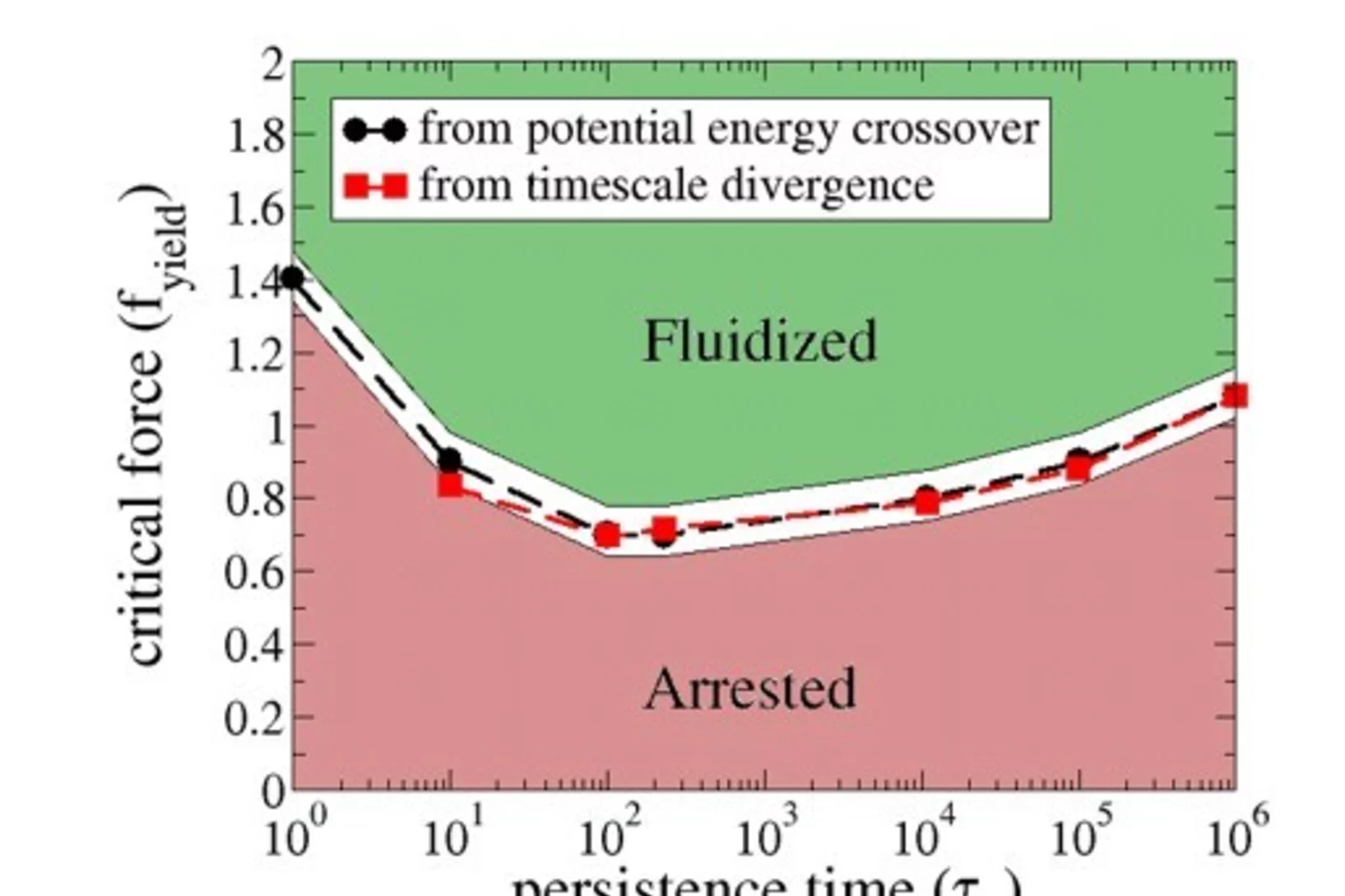
We computationally investigate the transition from rigid to flowing states in dense assemblies of self-propelled particles. Such theoretical representations of biological assemblies have yielded tremendous insight into collective behaviour across many scales, from bird flocks, through bacterial colonies, tissue organisation and including sub-cellular assemblies such as the cytoskeleton. Of particular interest to us are observations of dramatic changes in the dynamics of chromatin within cell nuclei, understood to undelie changes in biological state and function. Dynamics in this context is controlled by the strength and temporal persistence of out-of-equilibrium mechanical perturbations, as well as the geometry of confinement. Evidence of a transition from rigid to flowing states across a critical perturbation strength, strongly reminiscent of yielding in externally deformed amorphous solids, motivates us to explore this analogy, and to investigate the role of persistence time and confinement geometry on the transition.
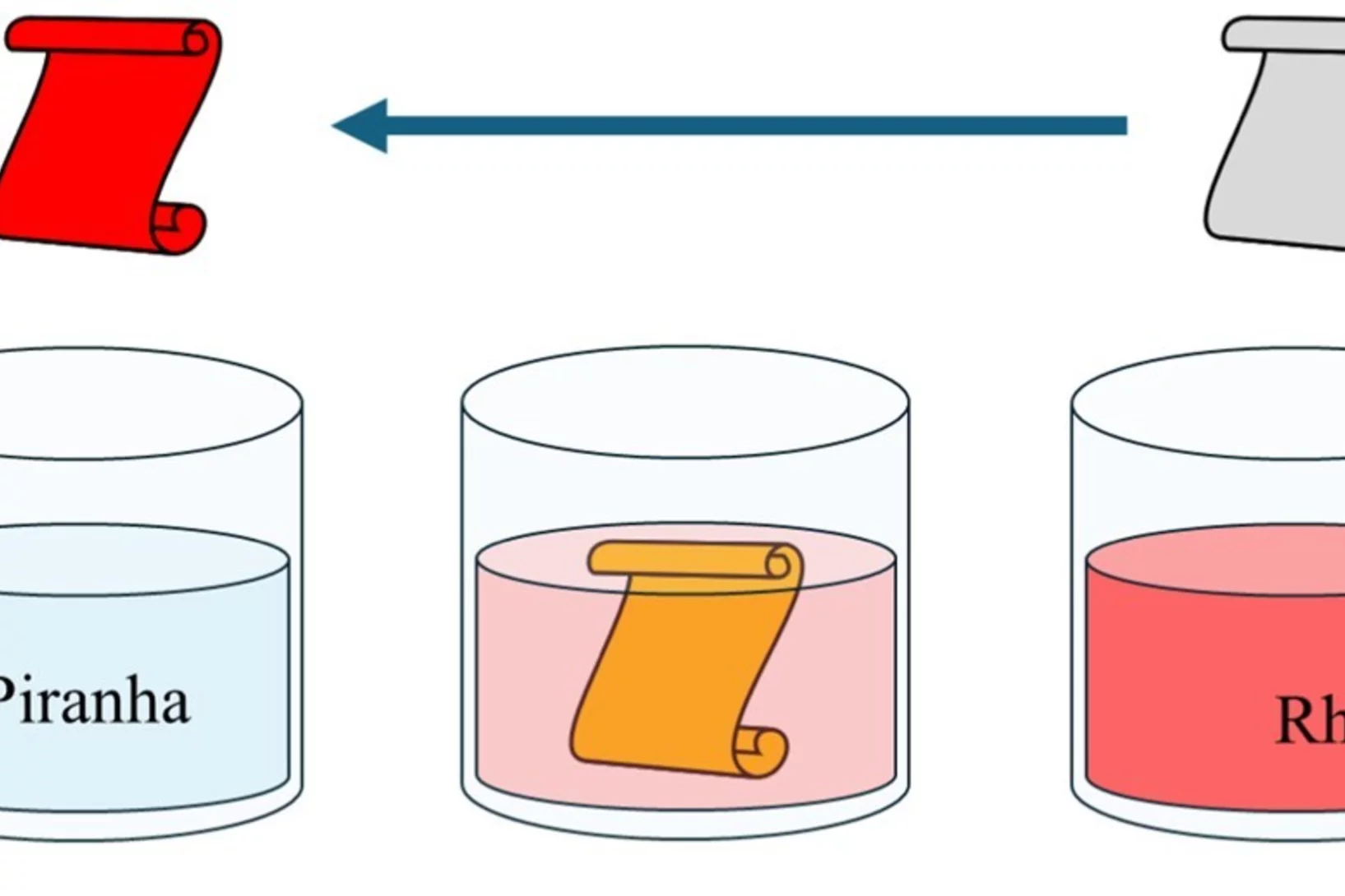
Rhodium recovery from acidic wastewater using radiografted chelating adsorbents
Platinum group metals (PGMs), particularly rhodium (Rh), are rare and vital for industrial applications, with Rh being scarcer (≈ 20 t/y) than platinum (Pt) and palladium (Pd). Its high cost and limited supply emphasize the need for efficient recovery from industrial waste. New radiografted chelating adsorbents, created through irradiation, offer a sustainable, cost-effective alternative to existing extraction methods. They exhibit high efficiency, selectivity, and reusability, making them ideal for recovering and recycling Rh from industrial wastewater.
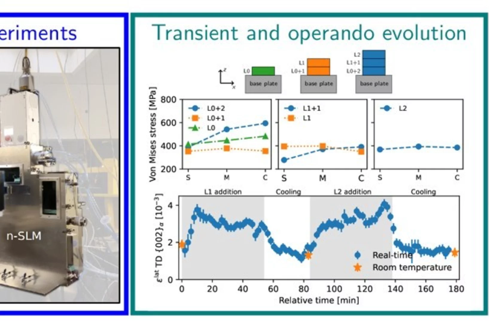
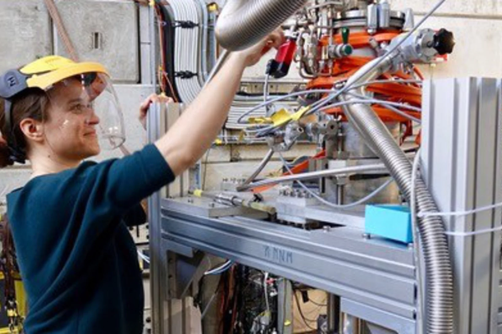
Evolution of texture and residual stresses in 2205 duplex stainless steel during laser powder bed fusion
This study uses a custom-designed laser powder bed fusion machine, capable of operating in neutron instruments, to track metallic material evolution during additive manufacturing process. More specifically, it investigates the development of residual stresses in textured 2205 duplex stainless steel during laser powder bed fusion. In situ and operando neutron diffraction experiments were conducted to study the transient and real-time evolution of stresses and strains during processing, using an AM machine designed for neutron studies. Additionally, Bragg-edge imaging was employed to investigate the crystallographic texture. The results showed that residual stress redistribution primarily occurs in the first set of added layers when further layers are added on top. The cube texture observed in the sample significantly affects residual stress determination, leading to inaccuracies up to 96 MPa if not accounted for. This highlights the need for orientation-dependent diffraction elastic constants in residual stress calculations. Furthermore, variations in texture intensity across the sample dimensions were found to be driven by changes in the local temperature history, which were deciphered from real-time strain measurements. Finally, this study demonstrates the potential of combining laser powder bed fusion with neutron diffraction to investigate the underlying mechanisms of additive manufacturing in the bulk of the sample.
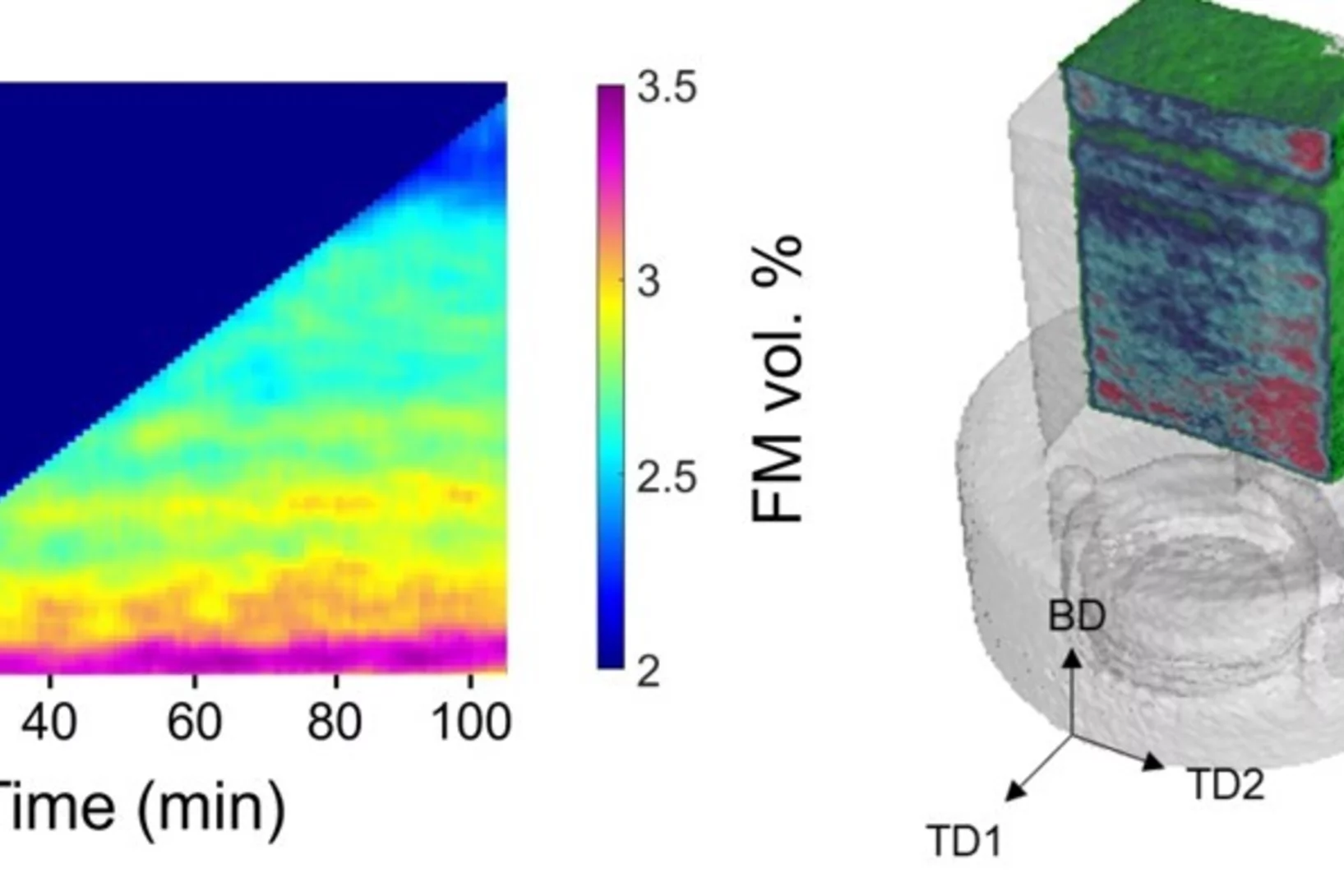
Operando phase mapping in multi-material laser powder bed fusion
This study introduces a custom-designed laser powder bed fusion machine, capable of operating in neutron instruments, to track material evolution during additive manufacturing. Using neutron imaging, this study uncovers how complex thermal cycling impacts phase development in multi-material parts, offering new insights into optimizing additive manufacturing processes and enhancing material properties for more precise and high-performance components.
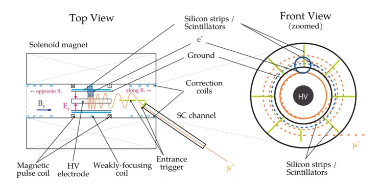
Anomalous spin precession systematic effects in the search for a muon EDM using the frozen-spin technique
In the paper, the international muEDM collaboration at PSI discusses systematic effects of the most sensitive measurement of the muon's electric dipole moment (EDM). Scientists from Europe are developing a prototype experiment using the frozen-spin technique (FST) to achieve unprecedented sensitivity. The FST meticulously aligns a magnetic field with a perpendicular electric field so that the muon's spin orientation always follows its momentum. This enhances the sensitivity to the muon EDM by about 3 orders of magnitude compared to the best result from the muon g-2 experiment at Brookhaven National Lab.
The paper addresses systematic effects that could mimic an EDM signal when E- and B-fields are not perfectly aligned, adjusted, or stable over time. While most effects cancel out when reversing the magnetic field, some residual effects the specifications for the fields' uniformity, stability, and orientation,
which are challenging but achievable.
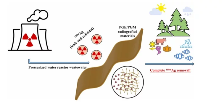
Removal of ionic and colloidal 110 mAg from radioactive wastewater using radiografted chelating adsorbents
Nuclear power plays a crucial role in a sustainable future due to its ability to generate large amounts of low-carbon electricity, which is essential for mitigating climate change. Unlike fossil fuels, nuclear energy produces minimal greenhouse gas emissions, helping to reduce the overall carbon footprint of power generation. However, the main concern is the inevitable accumulation of nuclear waste, and this needs to be properly addressed. With the anticipated increase in the number of operating nuclear power plants around the world it is essential to develop new materials and technologies for nuclear waste management. In our latest study we have developed and tested new radiografted materials as potential 110mAg adsorbents. This silver radionuclide is a very elusive contaminant in the pressurized water reactors (PWR) and represents a major problem for normal operation. Additionally, 110mAg possess a significant danger to the environment, if not removed completely from the PWR wastewater.
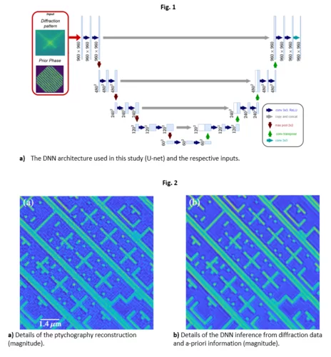
Towards fast ptychography image reconstruction of EUV masks by deep neural networks
In this study, we explore and demonstrate a rapid method for actinic patterned EUV mask inspection based on a deep neural network (DNN) architecture which exploits a-priori information of the photomask sample. We aim to achieve fast, high-quality image reconstruction of an EUV mask by using comparatively few diffraction patterns in a formalism consistent with the ptychography approach.
We tested our prior-primed DNN method on both synthetic and experimental data, demonstrating that the sample can be reconstructed fast and with high fidelity, allowing us to map out the mask defects down to a size of about 40 nm.
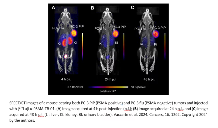
Design and Preclinical Evaluation of a Novel Prostate-Specific Membrane Antigen Radioligand Modified with a Transthyretin Binder
Radioligands targeting the prostate-specific membrane antigen (PSMA) are currently used in the clinics to treat patients with metastatic castration-resistant prostate cancer. Continuous investigations are, nevertheless, conducted to design new small molecule-based radioligands and improve their respective pharmacokinetic properties. Various strategies have been devised to reasonably prolong the blood circulation, which would result into enhanced tumor accumulation and radiation dose delivered to eliminate the cancer cells. The goal of this study was to investigate the influence of the incorporation of a transthyretin binder (TB-01) in the tumor uptake of the resultant PSMA-targeted radioligand.
Optimizing a radiochemical separation of 26Al from an acidic V-rich matrix
At the Paul Scherrer Institute (PSI), within the Isotope and Target Chemistry (ITC) group, various radiochemical methods are developed to fully separate and purify individual radionuclides. These separation methods are devised for both new experiments and for reprocessing radioactive waste from previous experiments.
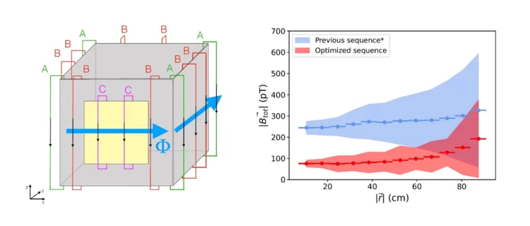
Achieving ultra-low and -uniform residual magnetic fields in a very large magnetically shielded room for fundamental physics experiments
n2EDM is the current state of the art experiment carrying out a high-precision search for an electric dipole moment of the neutron at the ultra-cold neutron source of PSI. In order to reach it’s incredible precision of 10-27 e cm, a stable and uniform magnetic environment is critical. Thus, shielding the experiment from external magnetic flux and preparing a pristine magnetic environment is crucial. To achieve this, n2EDM uses both passive and active magnetic shielding components. External, or residual, magnetic field contributions must be near-zero, and can be achieved via “degaussing” the experiment’s passive magnetic shielding. Degaussing reduces, ideally “erases”, the residual magnetization of a material. In this work, we greatly improved the degaussing procedure of n2EDM, reducing the residual magnetic field by a factor of two, improving its uniformity, and all while taking less time and dissipating less heat.
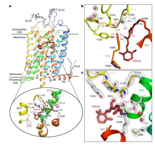
Ultrafast structural changes direct the first molecular events of vision
The visual pigment rhodopsin plays a critical role in the process of low-light vision in vertebrates. It is present in the disk membranes of rod cells in the retina and is responsible for transforming the absorption of light into a physiological signal. Rhodopsin has a unique structure that consists of seven transmembrane (TM) α-helices with an 11-cis retinal chromophore covalently bound to the Lysine sidechain of 7th TM helix. A negatively charged amino acid (glutamate) forms a salt bridge with the protonated Schiff base (PSB) of the chromophore to stabilize the receptor in the resting state.
Rhodopsin transforms the absorption of light into a physiological signal through conformational changes that activate the intracellular G protein transducin—a member of the Gi/o/t family—initiating a signaling cascade, resulting in electrical impulses sent to the brain and ultimately leading to visual perception. Although previous studies have provided valuable insights into the mechanism of signal transduction in rhodopsin, methods that provide both a high spatial and temporal resolution are necessary to fully understand the activation mechanism at the atomic scale from femtoseconds to milliseconds. This study presents the first experimentally-derived picture of the rhodopsin activation mechanism at the atomic scale using time-resolved serial femtosecond crystallography in association with hybrid quantum mechanics/molecular mechanics (QM/MM) simulations. The results show that light-induced structural changes in rhodopsin occur on a timescale of hundreds of femtoseconds, and they reveal new details about the conformational changes that occur during activation.
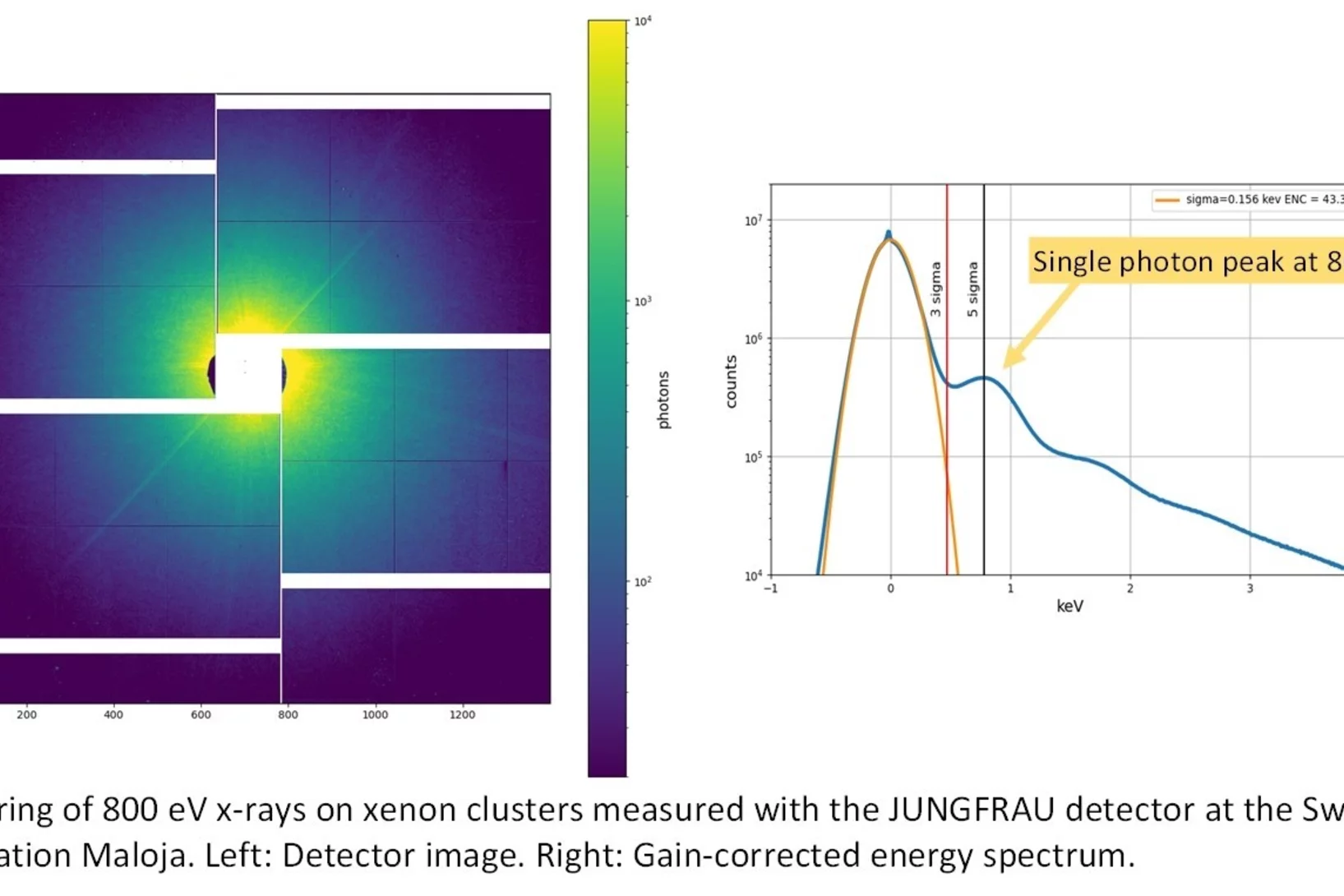
Advancing the JUNGFRAU detector toward low-energy X-ray applications
“Soft” x-rays are notoriously hard to detect. Particularly, in the context of high-performance synchrotron and free electron laser (FEL) experiments, suitable detector options for low-energy x-rays are highly sought after. Currently available options only provide limited area, readout speed, and dynamic range. Now, a team of scientists from the Laboratory for X-Ray Nanoscience and Technologies (LXN) at PSI are challenging these limitations. They combined a detector made at PSI with newly developed silicon sensors to push the resolution toward the soft x-ray limit. A first version of this detector system is now in operation at the SwissFEL endstation Maloja. And it points to further possibilities to refine the detector technology to eventually catch the elusive soft x-rays.
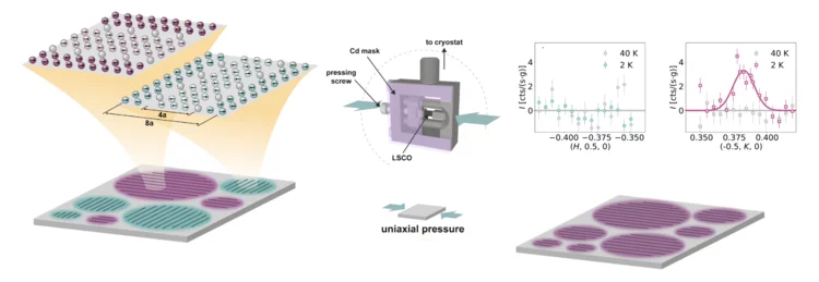
Single-domain stripe order in a high-temperature superconductor
The coupling of spin, charge and lattice degrees of freedom results in the emergence of novel states of matter across many classes of strongly correlated electron materials. A model example is unconventional superconductivity, which is widely believed to arise from the coupling of electrons via spin excitations. In cuprate high-temperature superconductors, the interplay of charge and spin degrees of freedom is also reflected in a zoo of charge and spin- density wave orders that are intertwined with superconductivity ...
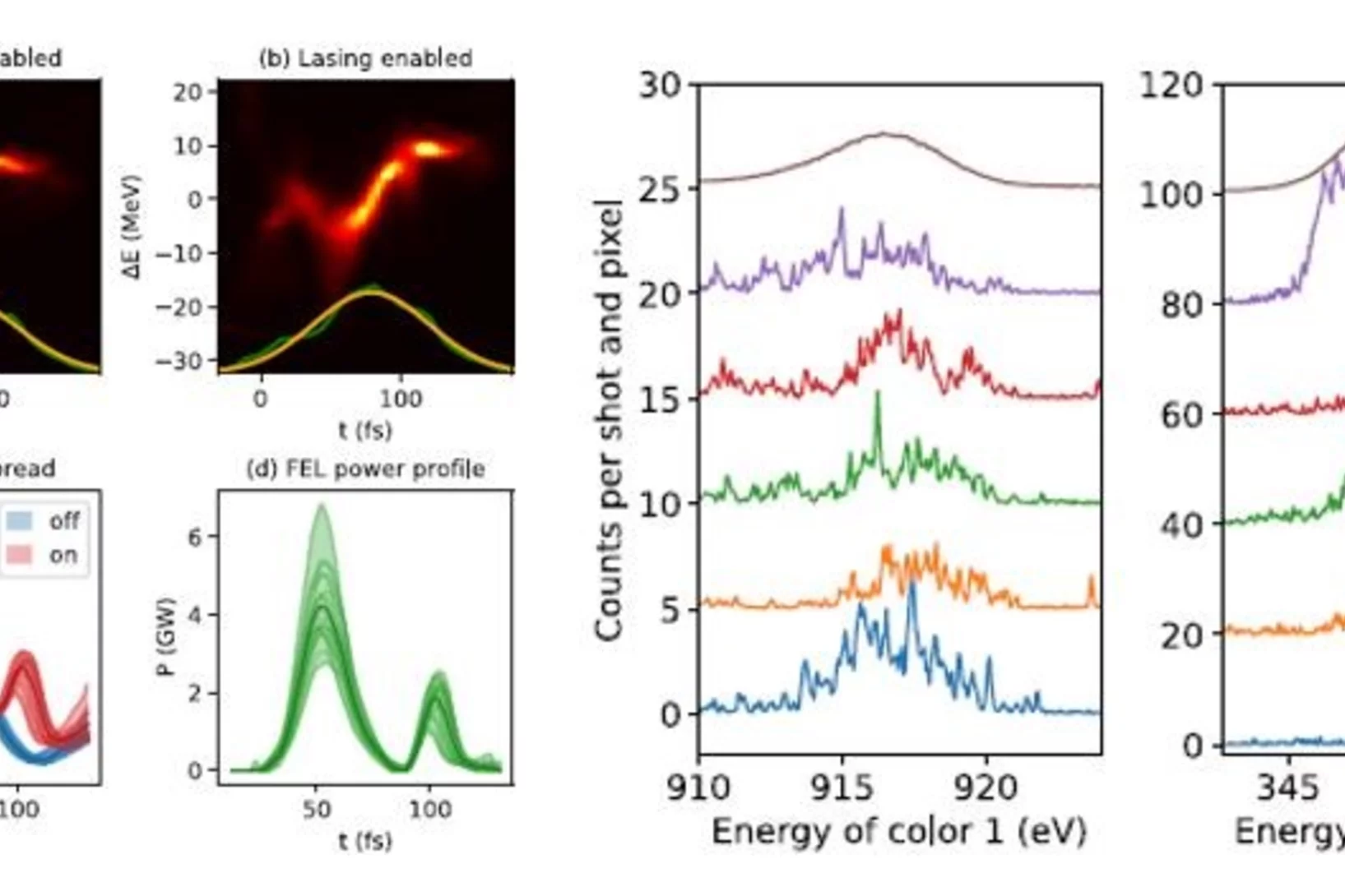
Widely tunable two-color x-ray free-electron laser pulses
SwissFEL team has demonstrated the generation of widely tunable two-color x-ray free-electron laser (FEL) pulses with unprecedented photon energy ratio between the two colors of about three (350 and 915 eV), in addition to a tunable time separation between the two pulses from negative time delays to up to 500 fs. These new capabilities open new opportunities to study ultrafast x-ray-induced energy transfer and relaxation processes in physics, chemistry, and biology.
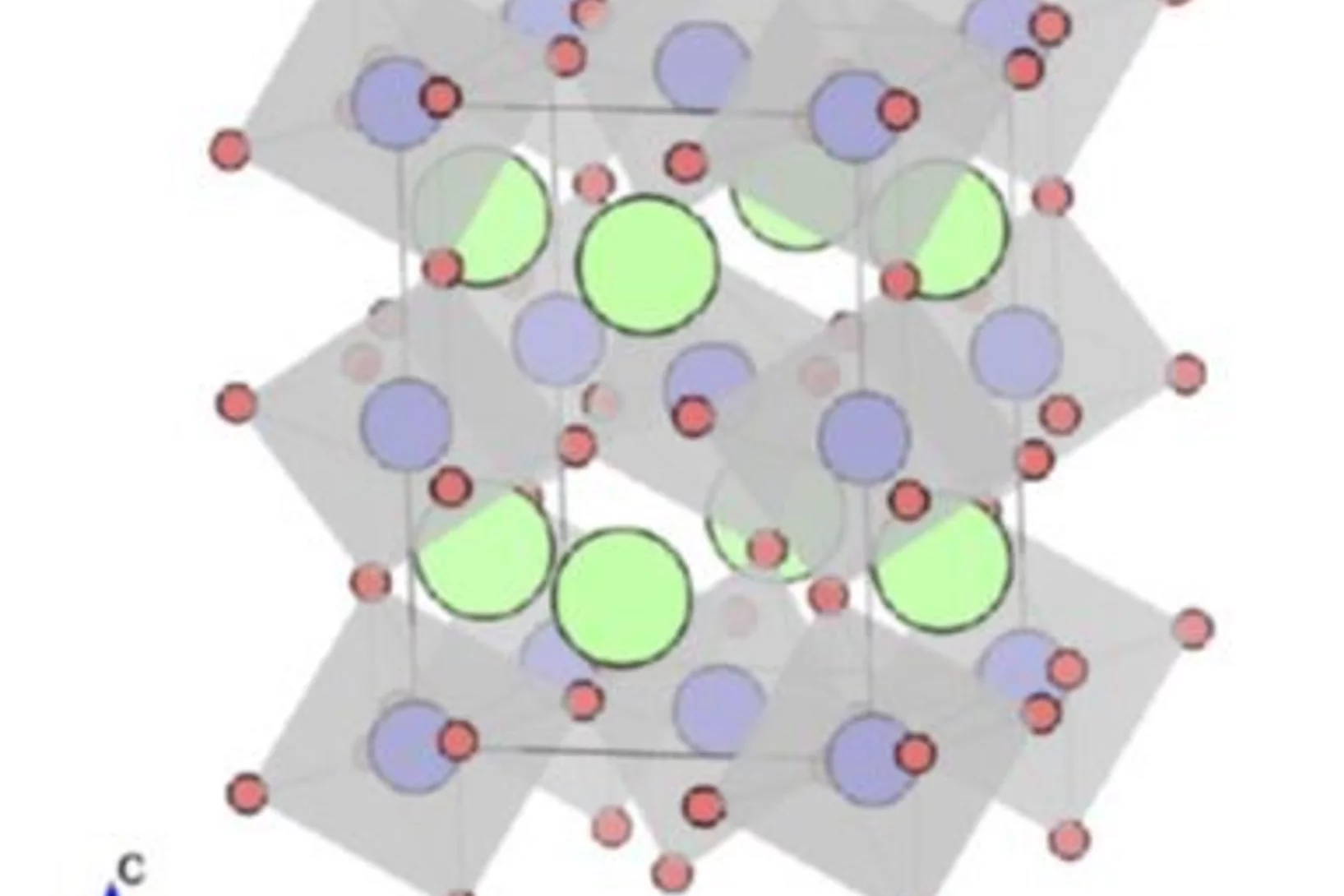
Low-energy spin dynamics in rare-earth perovskite oxides
A team of scientists from Paul Scherrer Institut and Oak Ridge National Laboratory review recent experimental studies of spin dynamics in the rare-earth perovskite materials. These compounds show unconventional magnetic excitations at low temperatures, including confined and deconfined spinons as well as multimagnon states, which were revealed by means of high-resolution neutron spectroscopy. These observations demonstrate that the rare-earth perovskite magnets can provide realizations of various aspects of quantum low-dimensional physics.
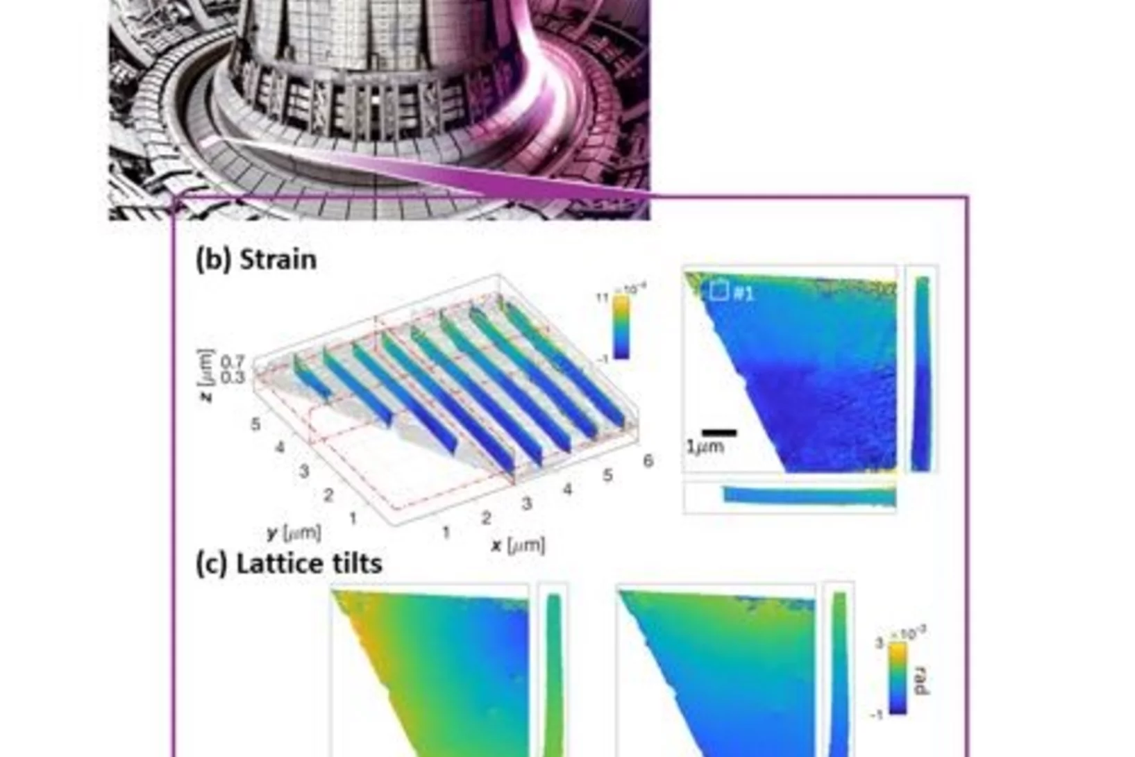
Revealing invisible defects in fusion reactor armor
In an exciting collaboration, Nick Phillips, a PSI Fellow at the cSAXS beamline, reveals nanoscale lattice distortions created by invisible defects in fusion reactor armor. This work develops the current understanding of how the smallest, but most prevalent defects, generated during neutron irradiation behave. The novel Bragg ptychographic approach published in Nature Communications paves the way for fast, robust, 3D Bragg ptychography.
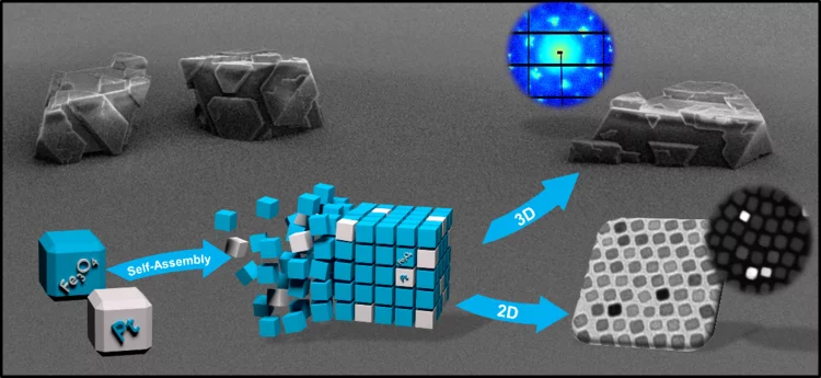
3D Binary Mesocrystals from Anisotropic Nanoparticles
A team of scientists from Konstanz have developed and characterized micrometre-size binary mesocrystals made from the self-assembly of ironoxide and platinum nanocubes and published their work in Angewandte Chemie International Edition. In collaboration with researchers from Empa and PSI, they used brilliant x-rays at the cSAXS beamline of the Swiss Light Source to characterize the lattice spacing in the crystalline structure of the mesocrystals and complement their results through electron microscopy.

Quantum billiards with correlated electrons
Our collaborators at the Jozef Stefan Institute – the leading author, Jan Ravnik, is now a PSI Fellow at LMN – report a study of the electron ordering in equilateral triangle structures via photoexcitation of the prototypical dichalcogenide 1T-TaS2.
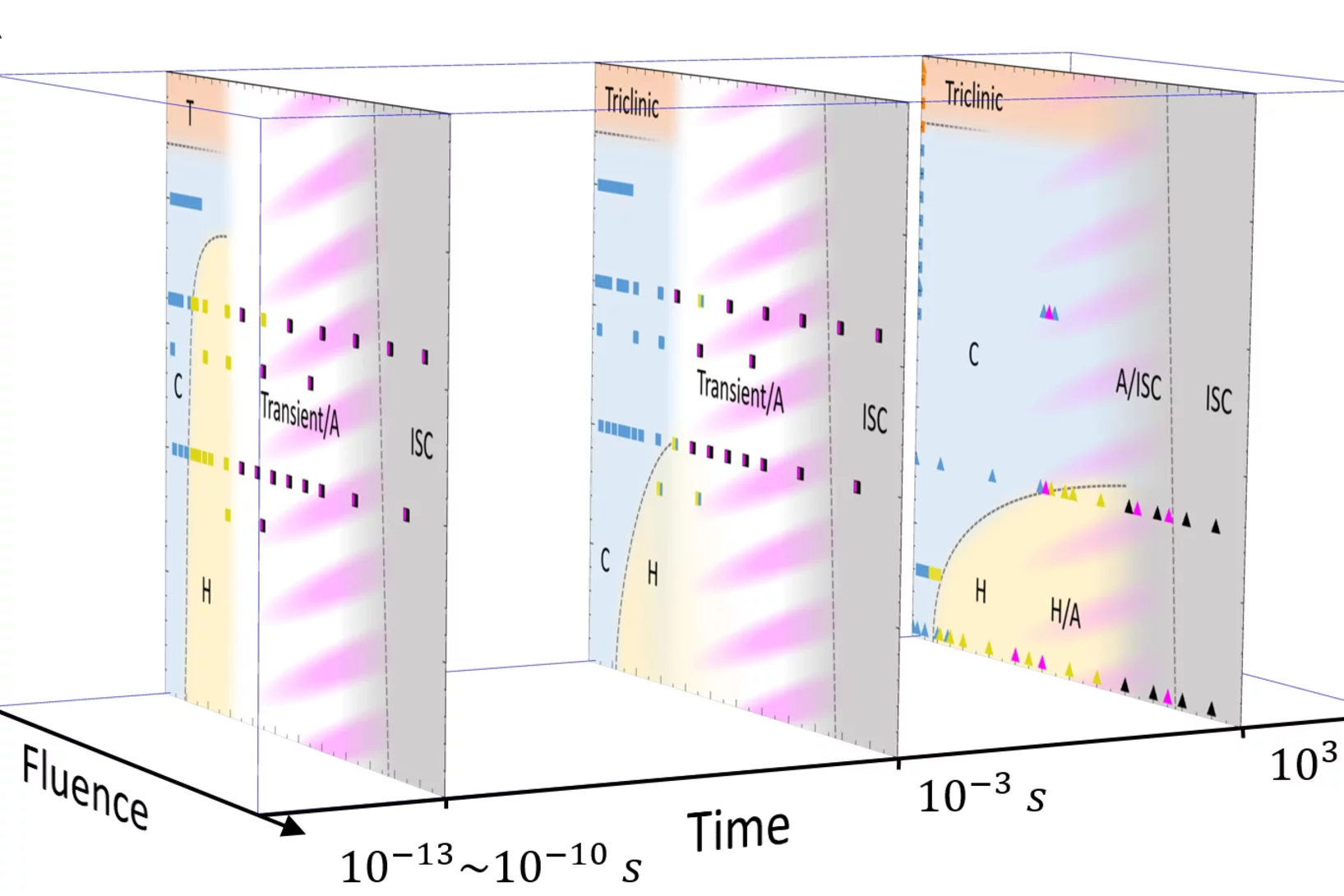
A time-domain phase diagram of metastable quantum states
Our collaborators at the Jozef Stefan Institute – the leading author, Jan Ravnik, is now a PSI Fellow at LMN – report a ‘dynamical’ phase diagram of metastable quantum states generated via photoexcitation of the prototypical dichalcogenide material 1T-TaS2.
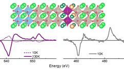
Hindering the magnetic dead layer in manganites
The authors demonstrate the stability of ferromagnetic order of one unit cell thick optimally doped manganite (La0.7Ba0.3MnO3, LBMO) epitaxially grown between two layers of SrRuO3 (SRO). LBMO shows ferromagnetism even above SRO Tc. Density Functional Theory calculations help understand the reasons behind this interesting result.

Looking inside airborne particles for the chemistry responsible for their adverse health effects.
Chemical changes inside of breathable airborne particles can cause reactive oxygen species (ROS) and carbon centered radicals (CCRs) to form, which are harmful to our bodies and induce oxidative stress in lungs. Using X-ray spectromicroscopy at the PolLux beamline and mimicking the environmental and sunlit conditions aerosol particles experience in the atmosphere near the Earth Surface, it was recently found that highly viscous organic particles with low water content can attain high concentrations of ROS and CCRs that persist over long times. Natural particles like these will occur in ambient humidity below 60% and effectively trap ROS and CCRs inside that react when exposed to light.
Split superconducting and time-reversal symmetry-breaking transitions in Sr2RuO4 under stress
Strontium ruthenate (Sr2RuO4) continues to present an important test of our understanding of unconventional superconductivity, because while its normal-state electronic structure is known with precision, its superconductivity remains unexplained. There is evidence that its order parameter is chiral, but reconciling this with recent observations of the spin part of the pairing requires an order parameter that is either finely tuned or implies a new form of pairing. Therefore, a definitive resolution of whether the superconductivity of Sr2RuO4 is chiral is important for the study of superconductivity.
Unconventional Transverse Transport above and below the Magnetic Transition Temperature in Weyl Semimetal EuCd2As2
As exemplified by the growing interest in the quantum anomalous Hall effect, the research on topology as an organizing principle of quantum matter is greatly enriched from the interplay with magnetism. In this vein, we present a combined electrical and thermoelectrical transport study on the magnetic Weyl semimetal EuCd2As2. Unconventional contribution to the anomalous Hall and anomalous Nernst effects were observed both above and below the magnetic transition temperature of EuCd2As2, indicating the existence of significant Berry curvature.
Ein elektronisches Material massschneidern
Forschende am PSI haben ein Material untersucht, das sich für zukünftige Anwendungen in der Datenspeicherung eignen könnte. Mit einem Trick haben sie die Kristallstruktur ihrer Probe gezielt verzerrt und dabei vermessen, wie dies die magnetischen und elektronischen Eigenschaften beeinflusst.

Strain engineering of the charge and spin-orbital interactions in Sr2IrO4
Understanding the relationship between entangled degrees of freedom (DOF) is a central problem in correlated materials and the possibility to influence their balance is promising toward realizing novel functionalities. In Sr2IrO4, the interaction between spin–orbit coupling and electron correlations induces an exotic ground state with magnetotransport properties promising for antiferromagnetic spintronics applications.
Mechanismus einer lichtgetriebenen Natriumpumpe aufgeklärt
Forschenden des Paul Scherrer Instituts PSI ist es erstmals gelungen, eine lichtgetriebene Natriumpumpe von Bakterienzellen in Aktion aufzunehmen. Die Erkenntnisse versprechen Fortschritte bei der Entwicklung von neuen Methoden in der Neurobiologie. Für ihre Untersuchungen nutzten die Forschenden den neuen Freie-Elektronen-Röntgenlaser SwissFEL.
Topological Magnetic Phase in the Candidate Weyl Semimetal CeAlGe
We report the discovery of topological magnetism in the candidate magnetic Weyl semimetal CeAlGe. Using neutron scattering we find this system to host several incommensurate, square-coordinated multi-k⃗ magnetic phases below TN. The topological properties of a phase stable at intermediate magnetic fields parallel to the c axis are suggested by observation of a topological Hall effect.
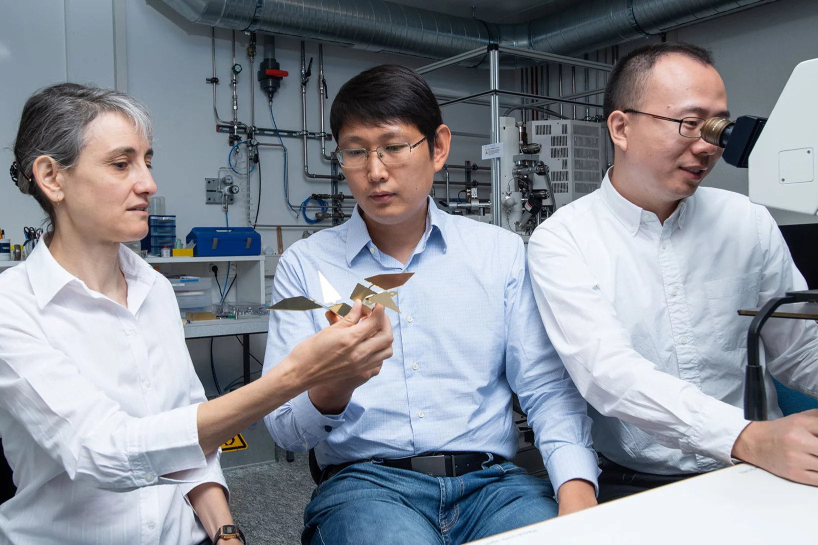
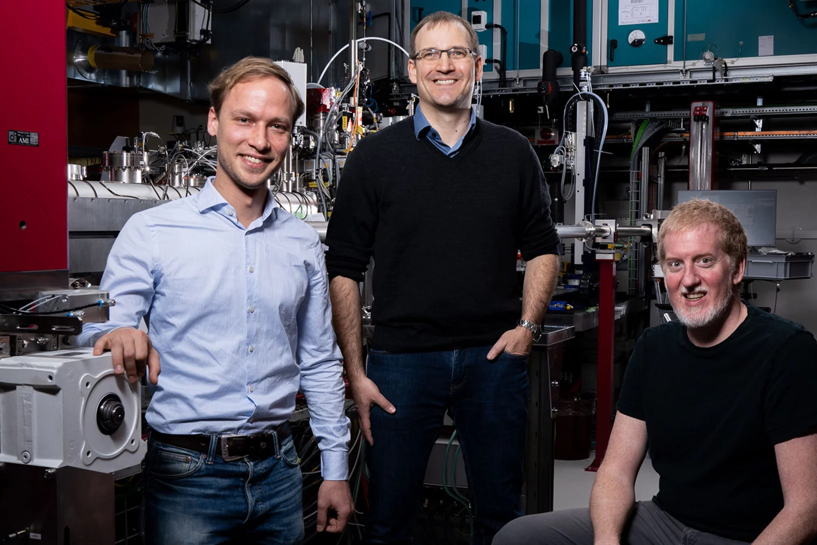
Auf dem Weg zu intelligenten Mikrorobotern
Er erinnert an einen Papiervogel, der mithilfe der japanischen Faltkunst Origami gefertigt wurde: ein Mikroroboter, der die Kraft von Magnetfeldern nutzt, um sich zu bewegen. Unter anderem in der Medizin könnten solche winzigen Maschinen bei Operationen zum Einsatz kommen.
Metastasierung von Tumoren verhindern
Forschende des Paul Scherrer Instituts PSI sind gemeinsam mit Kollegen des Pharmaunternehmens F. Hoffmann-La Roche AG der Entwicklung eines Wirkstoffes gegen die Metastasierung von bestimmten Krebsarten einen wichtigen Schritt nähergekommen. Mithilfe der Synchrotron Lichtquelle Schweiz SLS entschlüsselten sie die Struktur eines Rezeptors, der entscheidend an der Wanderung von Krebszellen beteiligt ist.