

Our research focuses on structure-based protein engineering to create biomolecules with novel functionalities
Our Research
Protein engineering is a powerful approach to generate biomolecules with novel functionalities. Fusion proteins can act as precise sensors to study changes in live cells, such as chemical changes, organelle movements, distances and involved forces. Engineered biomolecular nanomachines can capture viruses from fluids or perform other specific tasks. Fluorescent or electron-dense tags are important tools to track proteins in various imaging methods.
Proteins can be designed based on knowledge about the amino acid sequence and domain structure. Structure predictions using artificial intelligence-based algorithms play an important role in protein design, in addition to knowledge from experimental structural biology. Furthermore, a close look at the ultrastructure of natural proteins allows the identification of motifs and structures that can be built into designed proteins in new contexts (molecular biomimetics).
Our research focuses on structure-based protein engineering to create biomolecules with novel functionalities, with the following aims:
Design novel protein-based tags and sensors for multiscale imaging
A full understanding of biological processes requires knowledge about the dynamics and structures of biomolecules over multiple scales, from a molecular scale to a cellular scale and tissues. The structures of isolated proteins can be studied a near-atomic resolution using methods such as X-ray crystallography and cryo-electron microscopy (cryo-EM). The ultrastructure of cells can be tracked using light microscopy, including fluorescence microscopy and super-resolution microscopy. Important questions remain on the interplay between the molecular scale and the ultrastructure. The field of mesoscale structural biology (imaging and diffraction over a wide range of scales from near-atomic to cellular) requires novel tags, for example to optimally localize proteins of interest in correlative light- and electron-microscopy or to measure the dynamics of movements of components within live cells with a simple readout.
The design, engineering, production, characterization and application of such protein-based tags and sensors is an important aim on which we are focusing.
Engineer fusion proteins for the detection and inactivation of pathogens or diseased cells
We are constantly exposed to menaces originating from pathogens, including viruses, bacteria, fungi (and their toxins), prions, and viroids, as well as from defective cells such as cancer cells. The development of novel biological tools for the detection of pathogens and their interaction sites with the host contributes to an improved understanding of the molecular mechanisms underlying specific diseases. Furthermore, novel biological reagents hold promise for an improved, fast, and more reliable diagnosis.
Fusion proteins can comprise a simple or complex architecture. For example, a simple fluorescent protein-tagged receptor-binding domain of a pathogen can be useful to easily identify the corresponding receptor(s) on cultured cells. An engineered sensor that detects pathogen-induced changes in a cell is an example of a more complex fusion protein.
Our aim is to engineer fusion proteins that enable new possibilities for pathogen research and diagnosis, in the combat against new emerging diseases.
Related topics:
- https://www.psi.ch/en/psd/molecular-biology
- https://www.psi.ch/en/media/our-research/psi-advancing-in-the-fight-against-covid-19
- https://www.psi.ch/en/bio/covid-19
- https://www.psi.ch/en/media/our-research/how-botox-binds-to-neurons
- https://www.startupticker.ch/en/news/palto-therapeutics-enhances-rna-based-therapeutics
Optimize proteins for structural biology
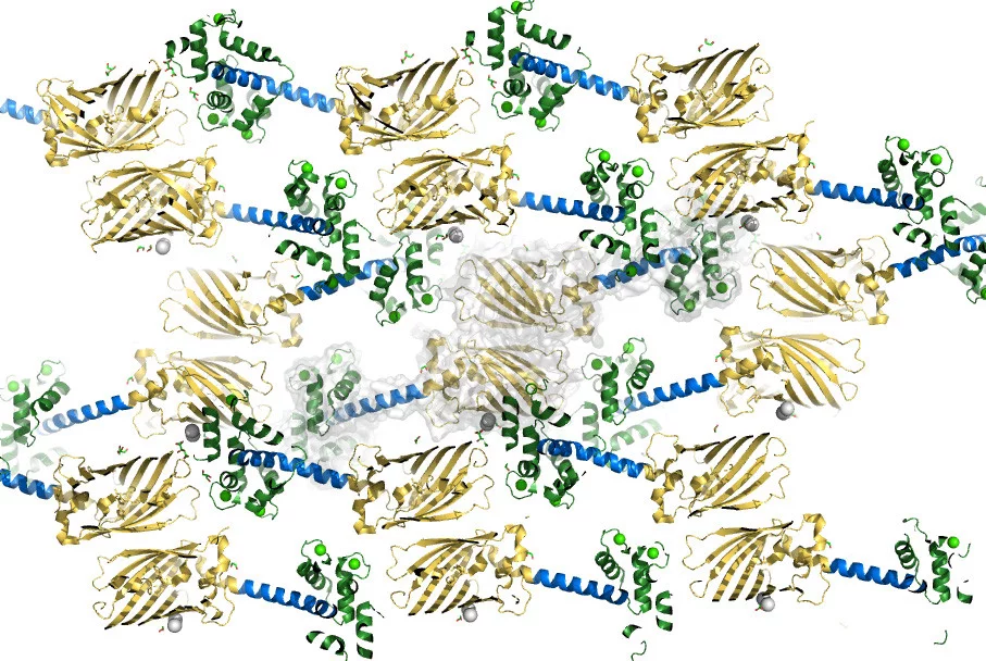
Dynamics and conformations are important factors in structural biology that can be studied by serial crystallography and cryo-EM. Sample quality is a main determinant for successful structure elucidation, and each method has its special sample requirements. For example, to study protein dynamics using X-ray free electron lasers (XFELs), the proteins of interest require conformational freedom within a crystal lattice. In cryo-EM, protein fusions that increase the mass of the protein of interest and add clearly identifiable features to aid angular orientation, without interfering with protein structure and function, can enable structure elucidation and/or improved resolution.
The integration of special linkers and spacers, inspired by natural proteins, allow the design and engineering of proteins that facilitate the detailed study of their natural function.
(Add Figure 4 later)
Related topics:
Selected References
(Since 2020)
-
Behbahanipour M, Benoit R, Navarro S, Ventura S
OligoBinders: bioengineered soluble amyloid-like nanoparticles to bind and neutralize SARS-CoV-2
ACS Applied Materials and Interfaces. 2023; 15(9): 11444-11457. https://doi.org/10.1021/acsami.2c18305
DORA PSI -
Farnung J, Muhar M, Liang JR, Tolmachova KA, Benoit RM, Corn JE, et al.
Semisynthetic LC3 probes for autophagy pathways reveal a noncanonical LC3 interacting region motif crucial for the enzymatic activity of human ATG3
ACS Central Science. 2023; 9(5): 1025-1034. https://doi.org/10.1021/acscentsci.3c00009
DORA PSI -
Collu G, Bierig T, Krebs A-S, Engilberge S, Varma N, Guixà-González R, et al.
Chimeric single α-helical domains as rigid fusion protein connections for protein nanotechnology and structural biology
Structure. 2022; 30(1): 95-106. https://doi.org/10.1016/j.str.2021.09.002
DORA PSI -
Bierig T, Collu G, Blanc A, Poghosyan E, Benoit RM
Design, expression, purification, and characterization of a YFP-tagged 2019-n CoV spike receptor-binding domain construct
Frontiers in Bioengineering and Biotechnology. 2020; 8: 618615 (10 pp.). https://doi.org/10.3389/fbioe.2020.618615
DORA PSI -
Skopintsev P, Ehrenberg D, Weinert T, James D, Kar RK, Johnson PJM, et al.
Femtosecond-to-millisecond structural changes in a light-driven sodium pump
Nature. 2020; 583: 314-318. https://doi.org/10.1038/s41586-020-2307-8
DORA PSI
Group members



Former team members
- Aristea Anna Leventi (Postdoctoral Researcher)
- Mara Wieser (Lab Technician)
- Daniel Abakumov (Intern)
- Dr. Gabriella Collu (PhD Student)
- Dr. Tobias Bierig (PhD Student)
- Dr. Anna-Sophia Krebs (Intern)
- Michaela Schertler (Intern)
- Niveditha Varma (Intern)