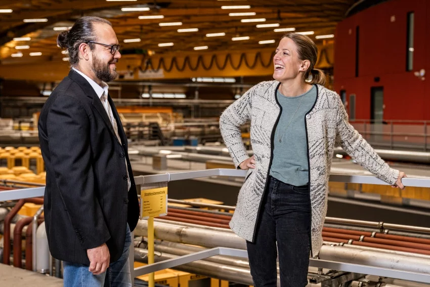
Two PSI researchers have won the 2021 Innovation Award on Synchrotron Radiation for 3D mapping of nanoscopic details in macroscopic specimens, such as bone. Marianne Liebi and Manuel Guizar-Sicairos are recognised for their “ground-breaking research and technology development enabling and implementing small-angle scattering tensor tomography.” They were awarded the international prize today by the Friends of Helmholtz-Zentrum Berlin (HZB).
Marianne Liebi is group leader of the Structure and Mechanics of Advanced Materials Group at PSI and tenure-track assistant professor at EPFL. Manuel Guizar-Sicairos is beamline scientist at the Swiss Light Source’s (SLS) cSAXS beamline.
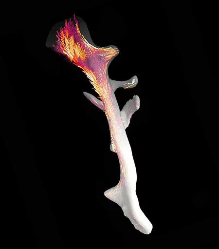
With their invention of small angle scattering tensor tomography (SASTT), work that they began at the SLS in 2013, Liebi and Guizar-Sicairos addressed a problem that had plagued the biological and material science communities: how to characterise nanostructures across macroscopic samples in 3D. At a single point, small angle scattering (SAXS) enables collection of information about the nanostructure of a sample - for example, the size and orientation of features. In many materials, not only the nanostructure itself, but also its variation on a macroscopic scale is crucial to the mechanical properties. This is the case in bone, where collagen fibres, orientated along the direction of the bone, are a key factor to determining bone strength. Until the development of SASTT, no available imaging techniques could provide the combination of length scales necessary to enable the imaging of nanoscale collagen fibres over a large sample of bone tissue.
When used with raster scanning, SAXS can be used to create a 2D survey of the nanostructure across a macroscopic specimen. Yet add in rotation of the sample to create a 3D SAXS tomogram, where each voxel contains 3D information on the nanostructure, and the data set - essentially 6D – is intimidatingly large. In a paper in Nature in 2015, Liebi and Guizar-Sicairos solved this incredible numerical problem. From over 1 million SAXS images, using optimisation techniques, they were able to reconstruct a 3D tomogram of a trabecular bone. The 3D tomogram contains information on the direction of the nanostructures, namely the constituent collagen fibres, contained within each voxel. Their acquisition and reconstruction method is applicable to any specimen, whether for biological or material sciences investigations.
Since they first published their technique of SASTT in Nature in 2015, Liebi and Guizar-Sicairos have been unerring in their commitment to further developing and promoting the technique, and making it accessible to non-expert users. Thanks to their efforts, SASTT is now implemented at beamlines not only at SLS, but also at Diamond, ESRF, DESY and at two synchrotrons in the US, in experiments that improve our understanding of materials with hierarchical structure, such as biological tissues.
The Innovation Award on Synchrotron Radiation is bestowed each year for an achievement that has “contributed significantly to the further development of techniques, methods or uses of synchrotron and FEL radiation”. Liebi and Guizar-Sicairos’ incredible dedication, both to solving a problem that had stumped researchers for years and to its implementation in the community, has been recognised today with this award.
Text: Paul Scherrer Institute / Miriam Arrell
Contact
Prof. Dr. Marianne Liebi
Group Leader, Structure and Mechanics of Advanced Materials Group
Paul Scherrer Institut
Forschungsstrasse 111
5232 Villigen PSI
Switzerland
Telephone: +41 56 310 44 38
E-Mail: marianne.liebi@psi.ch
Dr. Manuel Guizar-Sicairos
Beamline Scientist cSAXS
Paul Scherrer Institute
Forschungsstrasse 111
5232 Villigen PSI
Switzerland
Telephone:+41 56 310 34 09
E-Mail: manuel.guizar-sicairos@psi.ch
Further Information
Nanostructure surveys on macroscopic specimens by small-angle scattering tensor tomography
M. Liebi, M. Georgiadis, A. Menzel, P. Schneider, J. Kohlbrecher, O. Bunk and M. Guizar-Sicairos,
Nature 19 November 2015
DOI: 10.1038/nature16056


