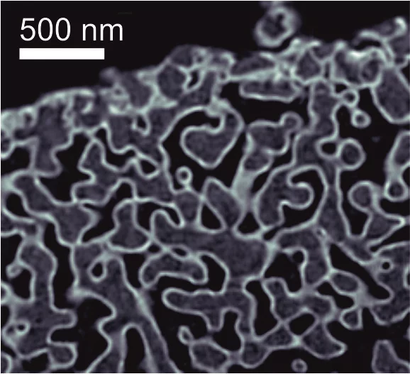
Tomographic microscopy has become an invaluable imaging method in both life and materials sciences. Oftentimes, high resolving power is required simultaneously with the ability to characterize large, statistically representative sample volumes. To this task, researchers at the Paul Scherrer Institut have established ptychographic computed tomography. The technique promises the ability to characterize three-dimensionally thousands of cubic microns with precision and resolution currently not achievable by other means. Using this technique, researchers at PSI now succeeded in demonstrating an isotropic 3D resolution of 16 nm, unmatched in X-ray tomography. They reported this first-of-its-kind demonstration on a 6 microns thick test object of Ta2O5-coated nanoporous glass in Scientific Reports.
The measurement was performed at the cSAXS beamline at the Swiss Light Source using a prototype instrument of the OMNY (tOMography Nano crYo) project. Whereas this prototype measures at room temperature and atmospheric pressure, the OMNY system, to be commissioned later this year, will provide a cryogenic sample environment in ultra-high vacuum without compromising imaging capabilities. The researchers believe that such a combination of ptychographic X-ray tomography with state-of-the-art instrumentation is a promising path to fill the resolution gap between electron microscopy and X-ray imaging, also in case of radiation-sensitive materials such as polymer structures and biological systems.
Read the full story
Reference
X-ray ptychographic computed tomography at 16 nm isotropic 3D resolutionM. Holler, A. Diaz, M. Guizar-Sicairos, P. Karvinen, Elina Färm, Emma Härkönen, Mikko Ritala, A. Menzel, J. Raabe & O. Bunk
Scientific Reports 4, Article number: 3857, DOI: 10.1038/srep03857
Contact
Dr. Mirko Holler, Swiss Light SourcePaul Scherrer Institute, 5232 Villigen PSI, Switzerland
Phone: +41 56 310 3613, e-mail: mirko.holler@psi.ch
