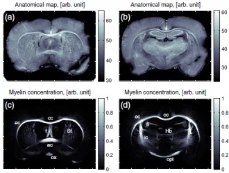
An international team of researchers from Denmark, Germany, Switzerland and France has developed a new method for making detailed X-ray images of brain tissue, which has been used to make the myelin sheaths of nerve fibres visible. Damage to these protective sheaths can lead to various disorders, such as multiple sclerosis. The facility for creating these images of the protective sheaths of nerve cells is being operated at the Swiss Light Source (SLS), at the Paul Scherrer Institute. The research team has reported on its work in the online version of the scientific journal NeuroImage.
Read the full story
Read the full story
Facility: SLS
T.H. Jensen, M. Bech, O. Bunk, A. Menzel, A. Bouchet, G. Le Duc, R. Feidenhans'l, F. Pfeiffer
NeuroImage, 2011;
DOI: 10.1016/j.neuroimage.2011.04.013
Paul Scherrer Institut
Phone: (+41) 56 310 3077, E-mail: oliver.bunk@psi.ch
Reference
Molecular X-ray computed tomography of myelin in a rat brainT.H. Jensen, M. Bech, O. Bunk, A. Menzel, A. Bouchet, G. Le Duc, R. Feidenhans'l, F. Pfeiffer
NeuroImage, 2011;
DOI: 10.1016/j.neuroimage.2011.04.013
Contact
Dr. Oliver BunkPaul Scherrer Institut
Phone: (+41) 56 310 3077, E-mail: oliver.bunk@psi.ch
