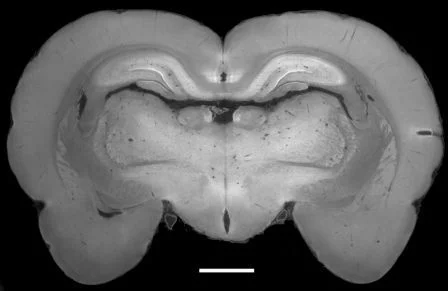
A new method forms the basis for the widespread use of an X-ray technique which distinguishing types of tissue that normally appear the same in conventional X-ray images
Traditional X-ray images can clearly distinguish between bones and soft tissue, with muscles, cartilage, tendons and soft-tissue tumours all look virtually identical. The phase-contrast technique developed a few years ago at the Paul Scherrer Institute enables X-ray images to be produced that clearly distinguish between these tissue types. Researchers at the Paul Scherrer Institute and the Chinese Academy of Science have now further developed the technique to such an extent that, in the future, it will be as simple to use as conventional X-rays. They anticipate that the process will help tumours to be detected in medical practices and could also help identify hazardous objects in luggage at airports. The researchers are reporting their findings this week in the online edition of the Proceedings of the National Academy of Sciences of the United States of America (PNAS).
Read full article
Facility: SLS
Swiss Light Source, Paul Scherrer Institut, 5232 Villigen PSI, Switzerland
Email: marco.stampanoni@psi.ch
Reference
Peiping Zhu, Kai Zhang, Zhili Wang, Yinjin Liu, Xiaosong Liu, Ziyu Wu, Samuel A. McDonald, Federica Marone, and Marco Stampanoni, PNAS Early Edition, 19 July 2010Contact
Prof. Dr. Marco StampanoniSwiss Light Source, Paul Scherrer Institut, 5232 Villigen PSI, Switzerland
Email: marco.stampanoni@psi.ch
