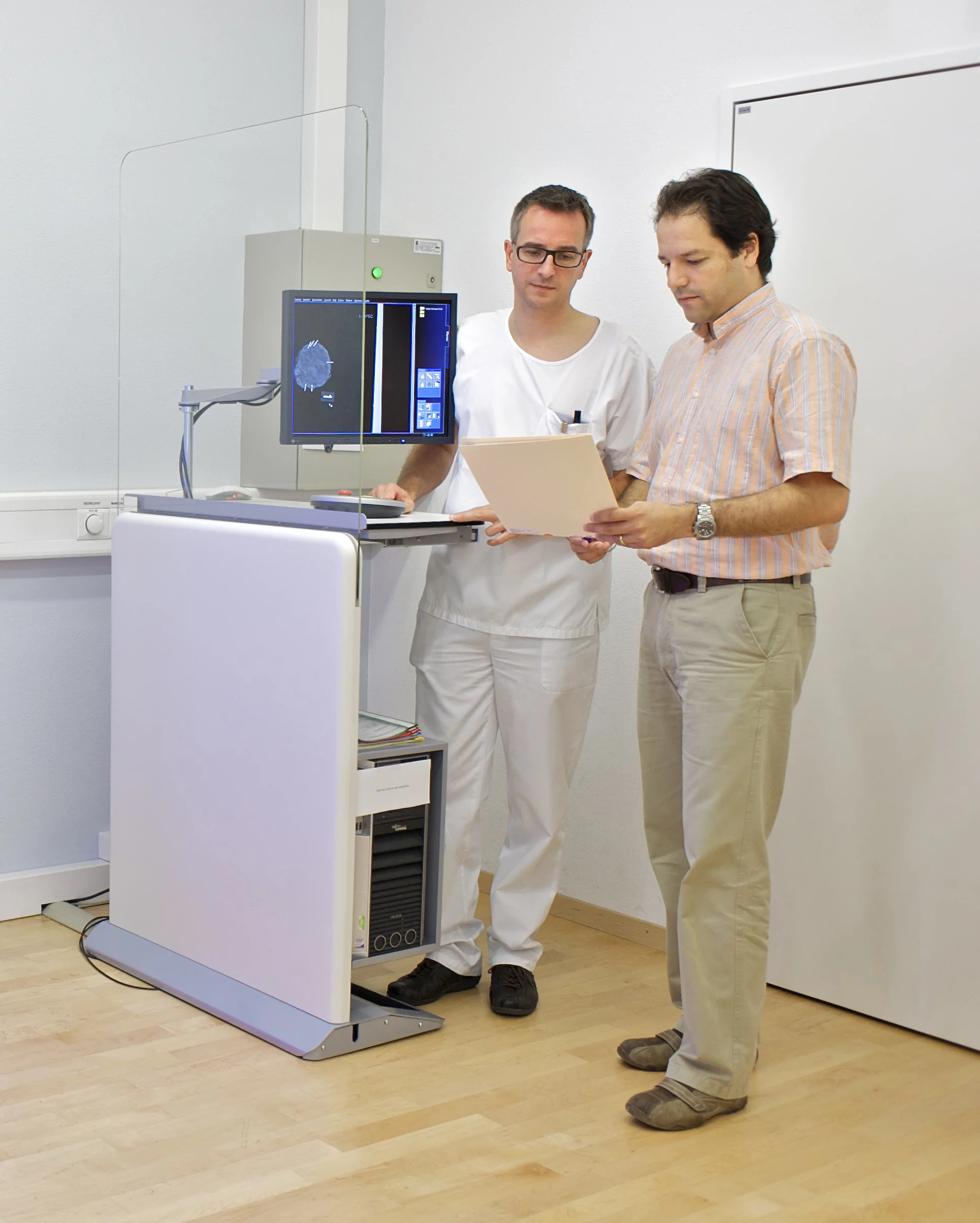
Collaboration between research, hospital and industry aimed at transferring innovative procedure into daily practice.
The Paul Scherrer Institute (PSI) has developed a new breast cancer diagnostic method, and is now carrying out first tests on non-preserved human tissue in conjunction with the Kantonsspital Baden AG. This new method should be able to reveal structures that cannot be seen using conventional mammography. Standard procedures only determine the extent to which X-rays are attenuated by various tissue structures. In contrast to this, the new method also makes use of the fact that X-rays actually consist of waves, and that their properties change slightly as they travel through tissue. These changes are now measurable and can contribute to the creation of a more meaningful image of the object under investigation. Scientists from the research department at Philips are currently investigating the use of this process as the basis for application in medical practice, and in mammography in particular. The researchers have reported on their results in the online edition of the
The aim of any mammography investigation is to detect tumours in the female breast as early as possible, so that treatment can start in good time. A good mammography procedure is therefore expected to recognise as many tissue changes as possible and to distinguish tumour tissue clearly from any other tissue. At the same time, the radiation dose administered during the investigation must be kept as low as possible.
Read the full story
Investigative Radiologyjournal.
The aim of any mammography investigation is to detect tumours in the female breast as early as possible, so that treatment can start in good time. A good mammography procedure is therefore expected to recognise as many tissue changes as possible and to distinguish tumour tissue clearly from any other tissue. At the same time, the radiation dose administered during the investigation must be kept as low as possible.
Read the full story
Facility: SLS
Stampanoni, Marco; Wang, Zhentian; Thüring, Thomas; David, Christian; Roessl, Ewald; Trippel, Mafalda; Kubik-Huch, Rahel A.; Singer, Gad; Hohl, Michael K.; Hauser, Nik
Investigative Radiology; published online 22 July 2011
DOI: 10.1097/RLI.0b013e31822a585f
Tel: +41 56 310 4724, +41 79 292 34 47; E-mail: marco.stampanoni@psi.ch
Dr. Nik Hauser, Chief Medical Officer at the Women’s Clinic, Certified Breast Centre, Kantonsspital Baden AG, CH-5404 Baden, Switzerland
Tel: +41 56 486 3636; E-mail: nik.hauser@ksb.ch
Reference
The First Analysis and Clinical Evaluation of Native Breast Tissue Using Differential Phase-Contrast MammographyStampanoni, Marco; Wang, Zhentian; Thüring, Thomas; David, Christian; Roessl, Ewald; Trippel, Mafalda; Kubik-Huch, Rahel A.; Singer, Gad; Hohl, Michael K.; Hauser, Nik
Investigative Radiology; published online 22 July 2011
DOI: 10.1097/RLI.0b013e31822a585f
Contact
Prof. Dr. Marco Stampanoni, Institute for Biomedical Engineering at ETH Zurich and the Laboratory for Macromolecules and Bioimaging at the Paul Scherrer Institute, 5232 Villigen PSI, SwitzerlandTel: +41 56 310 4724, +41 79 292 34 47; E-mail: marco.stampanoni@psi.ch
Dr. Nik Hauser, Chief Medical Officer at the Women’s Clinic, Certified Breast Centre, Kantonsspital Baden AG, CH-5404 Baden, Switzerland
Tel: +41 56 486 3636; E-mail: nik.hauser@ksb.ch
