• Paul Scherrer Institut PSI
  • PSI Research, Labs & User Services

Digital User Office

  • Digital User Office
  • DE
  • EN
  • FR
Paul Scherrer Institut (PSI)
Suche
Paul Scherrer Institut (PSI)

Hauptnavigation

  • Research at PSIOpen mainmenu item
    • Research Initiatives
    • Research Integrity
    • Scientific Highlights
    • Scientific Events
    • Scientific Career
    • PSI-FELLOW
    • PSI Data Policy
  • Research Divisions and LabsOpen mainmenu item
    • Overview
    • Research with Neutrons and Muons
    • Photon Science
    • Energy and Environment
    • Nuclear Energy and Safety
    • Biology and Chemistry
    • Large Research Facilities
  • Facilities and InstrumentsOpen mainmenu item
    • Overview
    • Large Research Facilities
    • Facilities
    • PSI Facility Newsletter
  • PSI User ServicesOpen mainmenu item
    • User Office
    • Methods at the PSI User Facilities
    • Proposals for beam time
    • Proposal Deadlines
    • Data Analysis Service (PSD)
    • EU support programmes
  • DE
  • EN
  • FR

Digital User Office (mobile)

  • Digital User Office

You are here:

  1. PSI Home
  2. Labs & User Services
  3. PSD
  4. LSB
  5. X-Ray Tomography
  6. Scientific Highlights
  7. Looking into the hints of early breast cancer detection

Secondary navigation

X-Ray Tomography Group

  • People
    • Alumni
  • Research
    • Synchrotron X-ray Tomography
    • Translational X-ray Imaging
    • X-ray Optics Design and Fabrication
  • Teaching and Education
    • Student Projects
  • Scientific Highlights
  • News
  • Publications
  • Download
30 July 2020

Looking into the hints of early breast cancer detection

microcalcs

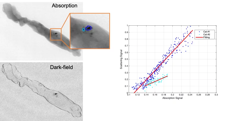
Microcalcifications consist of tiny calcium deposits, although they are usually benign, they are often present in areas of accelerated cell turnover, indicative for precancerous changes or early invasive breast cancer. Therefore, microcalcifications could act as indicators to tell whether an associated lesion is benign or malignant.  The ability to non-invasively discriminate between different microcalcifications can improve the diagnostic assessment by reducing the need for biopsies. The dark-field contrast provided by grating interferometry (GI) is sensitive to the microstructures of these tiny calcium deposits and reveal insights beyond the access of current mammography technology. The team of X-ray tomography group, in collaboration with Kantonsspital Baden, has carried out a reader study to investigative whether GI can discriminate benign and malignant microcalcifications and the results show the potential  of GI in non-invasively characterizing microcalcifications and aiding in the discrimination of benign from malignant lesions in clinical settings.

Original Publication

Can grating interferometry-based mammography discriminate benign from malignant microcalcifications in fresh biopsy samples?, Forte S, Wang Z, Arboleda C, Lång K,  Singer G, Kubik-Huch R  & Stampanoni M, European Journal of Radiology 129, 109077(2020). 

Contacts

Dr. Zhentian Wang 
Senior Scientist
Paul Scherrer Institut
Telephone: +41 56 310 5819
E-mail: zhentian.wang@psi.ch
 

Sidebar

top

Footer

Paul Scherrer Institut

Forschungsstrasse 111
5232 Villigen PSI
Switzerland

Telephone: +41 56 310 21 11
Telefax: +41 56 310 21 99

How to find us
Contact form

Visitor Centre psi forum
School Lab iLab (in German)
Center for Proton Therapy
PSI Education Centre
PSI Guest House
PSI Gastronomie (in German)


Service & Support

  • Phone Book/People Search
  • User Office
  • Accelerator Status
  • PSI Publications
  • Suppliers
  • E-Billing
  • Computing
  • Safety (in German)

Career

  • Working at PSI
  • Job Opportunities
  • Training and further education
  • Vocational Training (in German)
  • PSI Education Center

For the media

  • PSI in brief
  • Facts and Figures
  • Media Contact
  • Media Releases
  • Social Media Newsroom

Follow us: Twitter (in English) LinkedIn Youtube Issuu RSS

Footer legal

  • Imprint
  • Terms and Conditions
  • Editors' login