Breast cancer is the second leading cause of cancer-related deaths. The widely accepted life-saving countermeasure -- mammography -- is subject to criticism due to its limited sensitivity and specificity. One problem, the tissue overlap, especially problematic for women with dense breasts, is eliminated in a dedicated cone-beam breast CT. It does not, however, solve the other issue: the fact that the soft tissue of the breast provides limited contrast in an attenuation-based x-ray imaging.
The advent of highly efficient photon-counting detectors brought attenuation-based imaging to its physical limits. The contrast to noise ratio (CNR) can be only improved by increasing the dose. This limit, however, does not apply to phase-contrast imaging. In this method diffraction gratings with micrometre-scale lamellae imprint a pattern into the x-ray beam, which is then distorted by the refraction of x-rays in the object. Analysis of the distorted pattern yields in addition to attenuation images two additional ones: corresponding to a coherent refraction and to blurring by small, unresolvable structures. As the grating fabrication technology improves smaller and smaller refraction angles will be able to be detected increasing the contrast while keeping the dose same.
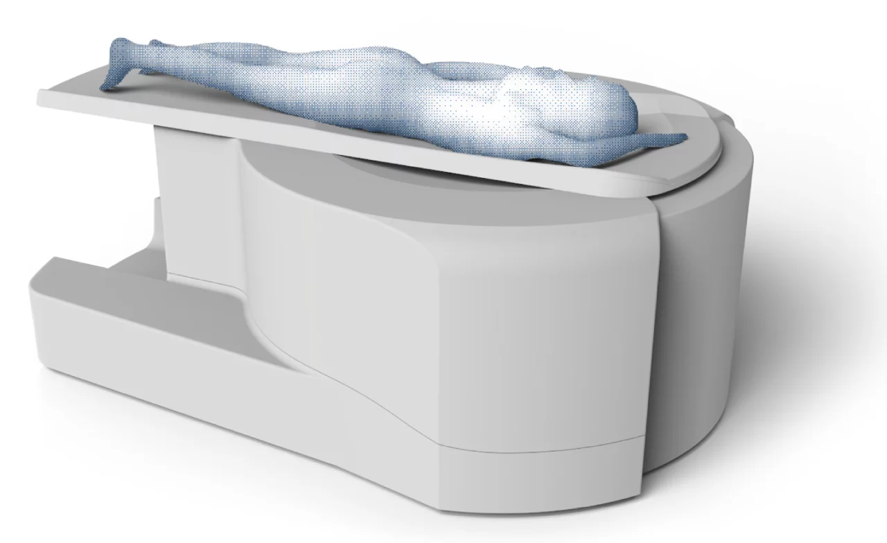
We are developing a large-field-of-view grating-based CT system, with parameters suitable for a clinical dedicated breast imaging. The design is a compact gantry rotating around a breast of a patient lying on a bed directly above it. We aim to prove the diagnostic value of phase contrast in breast cancer diagnostics. We collaborate with GratXray, a spin-off with roots in the group, on bringing the technology to clinics.
Collaboration
- GratXray, PARK innovAARE, 5234 Villigen, Switzerland
Funding
- This project is supported by Swisslos
Contacts
michal.rawlik@psi.ch
