• Paul Scherrer Institut PSI
  • PSI Research, Labs & User Services

Digital User Office

  • Digital User Office
  • DE
  • EN
  • FR
Paul Scherrer Institut (PSI)
Search
Paul Scherrer Institut (PSI)

Hauptnavigation

  • Research at PSIOpen mainmenu item
    • Research Initiatives
    • Ethics and Research integrity
    • Scientific Highlights
    • Scientific Events
    • Scientific Career
    • PSI-FELLOW
    • PSI Data Policy
  • Research Divisions and LabsOpen mainmenu item
    • Overview
    • Research with Neutrons and Muons
    • Photon Science
    • Energy and Environment
    • Nuclear Energy and Safety
    • Biology and Chemistry
    • Scientific Computing, Theory and Data
    • Large Research Facilities
  • Facilities and InstrumentsOpen mainmenu item
    • Overview
    • Large Research Facilities
    • Facilities
    • PSI Facility Newsletter
  • PSI User ServicesOpen mainmenu item
    • User Office
    • Methods at the PSI User Facilities
    • Proposals for beam time
    • Proposal Deadlines
    • Data Analysis Service (PSD)
    • EU support programmes
  • New ProjectsOpen mainmenu item
    • SLS 2.0
    • IMPACT
  • DE
  • EN
  • FR

Digital User Office (mobile)

  • Digital User Office

You are here:

  1. PSI Home
  2. Labs & User Services
  3. PSD
  4. SLS
  5. CSAXS
  6. Imaging strain with high resolution

Secondary navigation

CSAXS

  • Status
  • User Information
    • Application for beamtime
    • Operation Schedule
    • Manuals
    • Software
    • Infrastructure
  • People
  • Beamline Layout
    • Source
    • Optics
  • Endstations
  • Research
  • Publications
  • Scientific Highlights
  • Contacts
7 July 2021

Imaging strain with high resolution

A new method for strain imaging in crystalline materials, ptychographic topography, was developed by an international collaboration of researchers from the Paul Scherrer Institute (Switzerland), European XFEL (Germany), CNRS-University of Poitiers-ENSMA (France), ETH Zürich (Switzerland), and the Université Paris-Saclay (France). The method was developed at the cSAXS beamline, Swiss Light Source, and is available for user experiments. Ptychographic topography has important benefits compared to existing methods such as high resolution (tens of nanometers), ability to tackle complicated three-dimensional strain fields, simultaneous phase and amplitude information and the robustness of the reconstructions. The performance of the method was demonstrated on imaging strain in InSb micropillars after uniaxial compression with 29 nm spatial resolution.

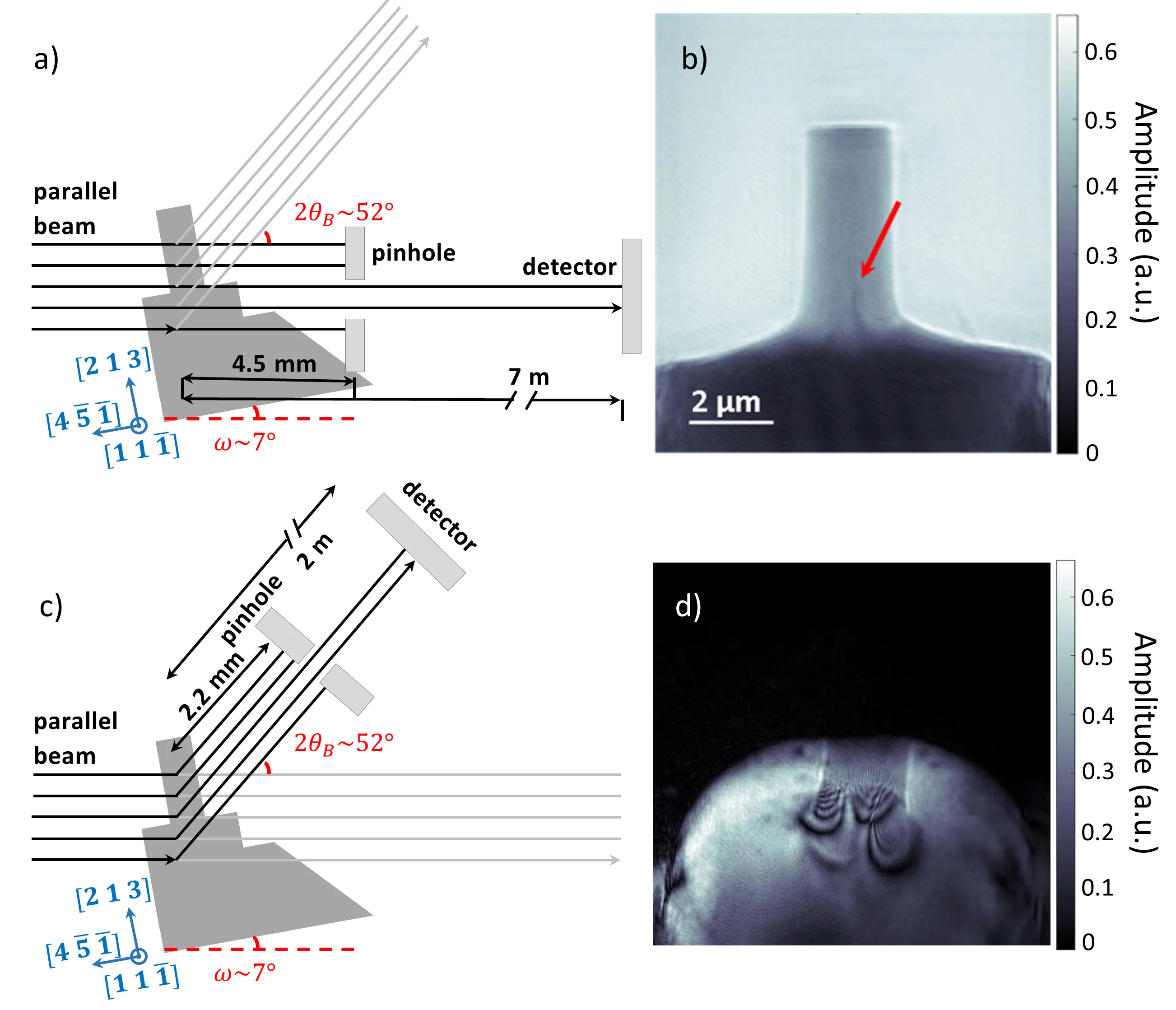
The method takes advantage of well-established X-ray topography and the idea of tele-ptychography. In ptychographic topography a crystal is placed in diffraction condition for specific crystallographic planes and ptychographic scans are performed in the forward (or diffraction) direction with a pinhole scanned several millimeters after the sample. In this way, it is possible to reconstruct the exit wave field at the pinhole position. The reconstruction, which corresponds to the wavefield at the plane of the pinhole, can be then numerically backpropagated to the crystal position (see Figure) and is sensitive to lattice imperfections.

Ptychographic topography provides high strain sensitivity, high resolution, and the flexibility needed to study complicated stain fields in extended specimens with an easy and robust reconstruction pipeline. The performance of the method will be soon boosted with upcoming upgrades of synchrotron storage rings. Most importantly, the simultaneous phase and amplitude reconstruction provided by this method opens a potential to retrieve quantitative three dimensional strain fields directly from the X-ray topography data.

Ptycho-topo figure
Ptychographic topography on InSb micropillars in (a) forward and (c) diffraction directions. (b) Amplitude of the image reconstruction obtained in the forward direction. Red arrow shows strained region on the image. (d) Amplitude of the image reconstruction in the diffraction direction.
Contact

Dr. Mariana Verezhak
Postdoctoral fellow, Swiss Light Source
Paul Scherrer Institut
Telephone: +41 56 310 32 53
E-mail: mariana.verezhak@psi.ch

Dr. Ana Diaz
Beamline scientist, Swiss Light Source
Paul Scherrer Institut
Telephone: +41 56 310 56 26
E-mail: ana.diaz@psi.ch

Dr. Steven Van Petegem
Senior Scientist, Laboratory for Condensed Matter (LSC)
Paul Scherrer Institut
Telephone: +41 56 310 25 37
E-mail: steven.vanpetegem@psi.ch

Original Publication

X-ray ptychographic topography: A robust nondestructive tool for strain imaging
Mariana Verezhak, Steven Van Petegem, Angel Rodriguez-Fernandez, Pierre Godard, Klaus Wakonig, Dmitry Karpov, Vincent L. R. Jacques, Andreas Menzel, Ludovic Thilly, and Ana Diaz
Phys. Rev. B 103, 144107 (2021). doi: 10.1103/PhysRevB.103.144107

Sidebar

top

Footer

Paul Scherrer Institut

Forschungsstrasse 111
5232 Villigen PSI
Switzerland

Telephone: +41 56 310 21 11
Telefax: +41 56 310 21 99

How to find us
Contact

Visitor Centre psi forum
School Lab iLab (in German)
Center for Proton Therapy
PSI Education Centre
PSI Guest House
PSI Gastronomie (in German)
psi forum shop

Service & Support

  • Phone Book
  • User Office
  • Accelerator Status
  • PSI Publications
  • Suppliers
  • E-Billing
  • Computing
  • Safety (in German)

Career

  • Working at PSI
  • Job Opportunities
  • Training and further education
  • Career Center
  • Vocational Training (in German)
  • PSI Education Center

For the media

  • PSI in brief
  • Facts and Figures
  • Media corner
  • Media Releases
  • Social Media

Follow us: Twitter (in English) LinkedIn Youtube Facebook Instagram Issuu RSS

Footer legal

  • Imprint
  • Terms and Conditions
  • Editors' login