The CXI group research focus is on the theoretical conception, implementation, and development of new imaging techniques anchored in mathematical modelling. We devise and advance computational X-ray nano-imaging methods and algorithms for fundamental and applied research.
We work in close collaboration with beamline scientists and domain scientists to develop methods and image reconstruction algorithms that harness capabilities of PSI’s large-scale facilities , such as the SLS and SwissFEL, combined with new synergistic collaborations with EPFL. The ultimate goal is to advance and develop computational X-ray microscopy for next-generation light sources. Below are some examples of lines of research of the group.
Ptychography, phase retrieval, and holography
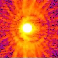
Coherent lensless imaging is in continuous development to circumvent limitations in quality and efficiency of conventional X-ray lenses. These methods use iterative algorithms to recover the wavefield phase, based on measurements of the corresponding diffracted intensity distribution. The intensity fringes and speckle are used for nanoscale imaging with the best resolution. We investigate and develop reconstruction pipelines, measurement schemes, and reconstruction algorithms to further the development and application of this technique in 2D and in 3D. More information.
Sparse hyperdimensional imaging
We apply concepts of sparsity to extend 3D measurements into the spectral and/or time domain. By leveraging the strong correlations in the multidimensional datasets the number of measurements can be greatly relaxed. As an example, here we extend nanoscale imaging with 28 nm 3D resolution to the spectral domain, which allows us to distinguish the enabling chemical contrast. Leveraging sparsity we used only 11% of the measurements that would be needed with conventional methods. Press release
Vectorial and tensorial tomography
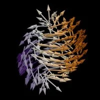
Under the right conditions X-rays can probe more than just a scalar property of the material, such as absorption or phase. For example, with X-ray circular magnetic dichroism the circularly polarized photons can probe one component of the magnetization vector. We develop measurement protocols and reconstruction algorithms that allow us to reconstruct from such measurement the full magnetization vector in each voxel, therewith revealing 3D nanoscale magnetization vortices, singularities (as shown), and general textures. For the case of small-angle X-ray scattering imaging, the voxel response can be modeled by a tensor. In that case measurements from millions of scattering patterns, each with a few million pixels, at different positions and orientations of the sample, are synthesized into such tensorial-tomograms in order to characterize the underlying 3D nanostructure. SAXS tensor tomography, Vector magnetization tomography


