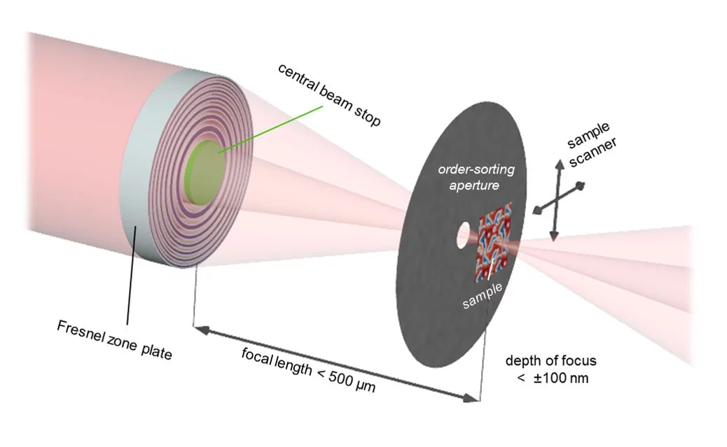
The short wavelengths of X-rays promise to reach spatial resolutions in the deep single-digit nanometer regime, providing unprecedented access to physical phenomena, such as magnetism at fundamental length scales. Despite considerable efforts in soft X-ray microscopy techniques, demonstrating a two-dimensional resolution beyond ten nanometers has not long remained a challenge for both scanning and full-field X-ray microscopy.
The developments in lithography-based diffractive optics by the X-ray Optics and Application group at PSI resulted in Fresnel zone plate lenses with structural sizes that can be as small as 6.4 nm [Rösner et al., Microsc. Microanal. 24 (Suppl. 2), 2018]. Utilising these lenses to their full capability is challenging in a number of ways. Firstly, the short focal length provides little space for blocking the unfocused, zero-order part of the transmitted X-ray beam. Secondly, very high mechanical precision is required to keep the sample in focus, and most importantly, to control the lateral positioning of sample relative to the focused X-ray beam. A critical part of the microscope system is the interferometric measurement of the sample position by laser beams in three dimensions and its coupling to the movement stages in order to overcome external vibrations. Implementing these lenses together with stability and precision improvements at the PolLux and HERMES scanning X-ray microscopes and a careful design of the experimental geometry, made the imaging of seven nanometer-sized structures possible. By combining this highly precise microscopy technique with the X-ray magnetic circular dichroism effect, the team reveals dimensionality effects in an ensemble of interacting magnetic nanoparticles.
In the recently published paper, PSI researchers and an international team of collaborators report on this achievement. The achieved imaging resolution of seven nanometers has never before been achieved in full-field or scanning soft X-ray microscopy, and is even competitive with coherent imaging techniques, such as ptychography.
Contacts
Benedikt Rösner (Principal Investigator)
Laboratory for Advanced Photonics
Paul Scherrer Institut, Forschungstrasse 111, 5232 Villigen PSI, Switzerland
Telephone: +41 56 310 2454, email: benedikt.roesner@psi.ch [English, German]
Christian David (high-resolution Fresnel zone plates)
Laboratory for Micro- and Nanotechnology
Paul Scherrer Institut, Forschungstrasse 111, 5232 Villigen PSI, Switzerland
Telephone: +41 56 310 3753, email: christian.david@psi.ch [English, German]
Armin Kleibert (magnetic nanoparticles)
Laboratory for Condensed Matter
Paul Scherrer Institut, Forschungstrasse 111, 5232 Villigen PSI, Switzerland
Telephone: +41 56 310 5527, email: armin.kleibert@psi.ch [English, German]
Original Publication
Soft x-ray microscopy with 7 nm resolution
Benedikt Rösner, Simone Finizio, Frieder Koch, Florian Döring, Vitaliy Guzenko, Manuel Langer, Eugenie Kirk, Benjamin Watts, Markus Meyer, Joshua Lorona Ornelas, Andreas spaeth, Stefan Stanescu, Sufal Swaraj, Rachid Belkhou, Takashi Ishikawa, Thomas Keller, Boris Gross, Martino Poggio, Rainer Fink, Jörg Raabe, Armin Kleibert, and Christian David
Optica 7, 2020, pp. 1602-1608


