An up-to-date publication list can be found in our repository DORA.
Biography
Benedikt Rösner is scientist in the Beamline Optics group at the Paul Scherrer Institute. He studied chemistry at the FAU Erlangen-Nürnberg, where he also obtained his PhD in Physical Chemistry. His dissertation focused on spectroscopic and microscopic characterization of electrically switchable metal-organic and organic materials. In 2016, he joined the X-ray Optics and Applications group at PSI as postdoctoral researcher, where he focused on the fabrication and application of transmissive diffractive optics. His main research goals where the fabrication of lenses for high-resolution X-ray microscopy, where 7 nm spatial resolution were achieved, the design and application of dedicated optical elements for experiments at X-ray free electron lasers, and the possibility to tailor beam properties via the design of optical elements. Since August 2019, he works in the Beamline Optics Group at the Swiss Light Source, where he is responsible for the upgrade of beamline optics for SLS 2.0.
Institutional Responsibilities
Benedikt Rösner's responsibility is the planning of beamline optics upgrades with respect to the storage ring upgrade to SLS 2.0. His main tasks are the evaluation of the changes on the accelerator side and their impact on beamline performance, the assessment of different beamline upgrade scenarios, techncial support and coordination of optics procurement, and the strategic procurement of optical elements that are used at several beamlines. In addition, he supports the SLS 2.0 project office with coordinative tasks regarding technical aspects as well as the project management within the Photon Science Division.
Scientific Research
World Record: Soft X-ray Microscopy with 7 nm Resolution
Fresnel zone plates are diffractive lenses widely used in X-ray microscopy. As an intrinsic limit from diffraction, their resolution is approximately in the order of their outermost zone width. This induces a major challenge in nanofabrication to produce smaller and smaller nanostructures striving for better resolution. In recent years, zone dimensions and resolution in X-ray micrscopy have been approaching the 10 nanometer level.
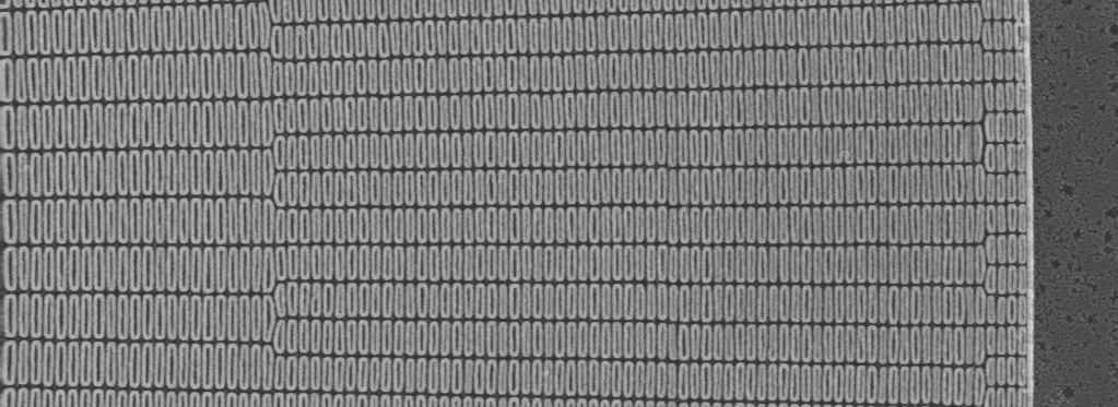
While zone plates have been reported on with focal spots well below 10 nm (cp. Mohacsi et al., Sci. Rep. 2017, Döring et al., Opt. Express 2013) reconstructed from scattering patterns in the far field, directly recorded X-ray micrographs have not been obtained so far. We thus fabricated zone plates with iridium zones with 9 nm (see above), and tested their resolution at the Hermes beamline at Soleil and the Pollux beamline at the Swiss Light Source. Indeed, these zone plates prove to be capable of resolving test structures with typical sizes down to 7 nm. The results were published in the OSA flagship journal OPTICA.
Light-Matter Interaction with OAM Beams
Creation of Optical Vortices
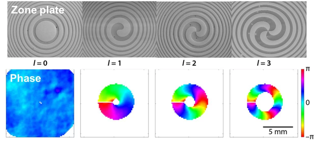
Photons have fixed spin and unbounded orbital angular momentum (OAM). A light wave, which carries an orbital angular momentum can be imagined as an optical vortex. While the spin momentum is manifested in the polarization of the electromagnetic field, the OAM corresponds to the spatial phase distribution of its wave front. The electric and magnetic field components of light can rotate uniformly clockwise or counterclockwise with respect to the light propagation, resulting in circular polarization. In optical vortices, it is the phase of the electromagnetic field that rotates around a circularity in a helical fashion. The vortex can be characterized with an integer-numbered topologic charge, which describes how often the wavefront is shifted around 360° within the distance of its wavelength.
To demonstrate optical vortices at free electron lasers, we fabricated spiral zone plates, which yield a diffraction pattern with such a phase singularity. The material of choice for the extremely intense EUV radiation of the FERMI free electron laser is silicon. We thus etched spiral zone plates into ultraflat thin silicon membranes, and characterized the radiation using a Hartmann wavefront sensor in the far field.
Viewpoint in APS Physics :: Wavefront Characterization of Optical Vortices - Phys. Rev. X
Photoelectric Effect with Transfer of the Optical Angular Momentum
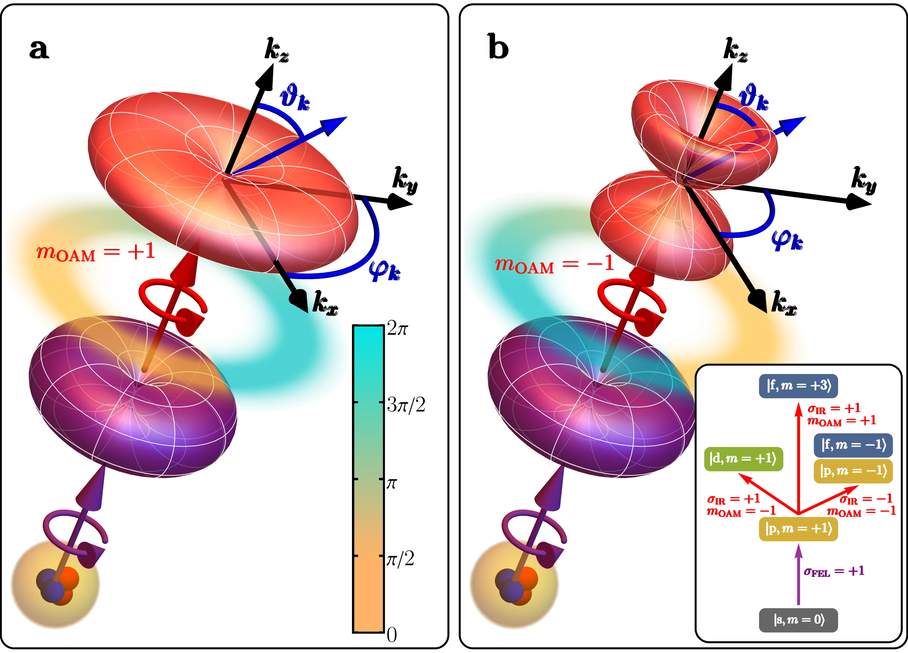
The distinctive way in which the photon spin dictates the electron motion upon light-matter interaction is the basis for numerous well-established spectroscopies that reveal the electronic, magnetic and structural properties of matter. In contrast, imprinting OAM onto a matter wave, specifically on a propagating electron, is generally considered very challenging and the anticipated effect undetectable. Indeed, this amounts to transferring the phase of a classical electromagnetic wave, defined within several hundreds of nanometres, to a quantum particle localized within the few angstroms of an atom. In addition, the centre of symmetry of irradiated atoms does not in general coincide with the axis of the photon beam.
In an experiment at the FERMI free electron laser, we demonstrate for the first time that OAM-based dichroism can be observed in photoelectron spectroscopy, using an extended sample of He atoms. Surprisingly, we find experimentally, and confirm theoretically, that the OAM of an optical field can be imprinted coherently onto a propagating electron wave, and that this phase information survives ensemble averaging out to macroscopic distances, where the electron is detected. We also show that electronic transitions, which are otherwise optically inaccessible due to selection rules, are essential for this process to occur. Our results reveal new aspects of light-matter interaction and point to a new kind of single-photon electron spectroscopy for accessing electronic optical transitions that are usually forbidden by symmetry. The results were published in Nature Photonics. A follow-up publication sheds light on the light-matter interaction and the possibility to induce magnetism. This paper represents the cover page article in Physical Review Letters Vol. 128/15 in April 2022.
Helical Dichroism
In addtion to the transfer of the OAM to free (photo-) electrons, an interesting question is whether the OAM can be transferred to electrons that are bound in a molecular orbital or in a band structure. In a row of experiments, we investigate the interaction of OAM beams with localized electrons, and dichroic effects when the direction of the optical vortex is reversed. For instance, we scanned magnetic vortices to see whether there is an intrinsic difference between the scattering pattern in dependence on the optical vortex. In other experiments, we perform X-ray absorption spectroscopy on chiral organometallic complexes, or on chiral organic molecules at various absorption edges. The results - published in Nature Photonics - indicate that there is indeed a strong dichrioc effect that is caused by the direction of the phase vortex, which has an exactly opposite sign when the enantiomer is changed to its mirror image.
X-ray transient gratings
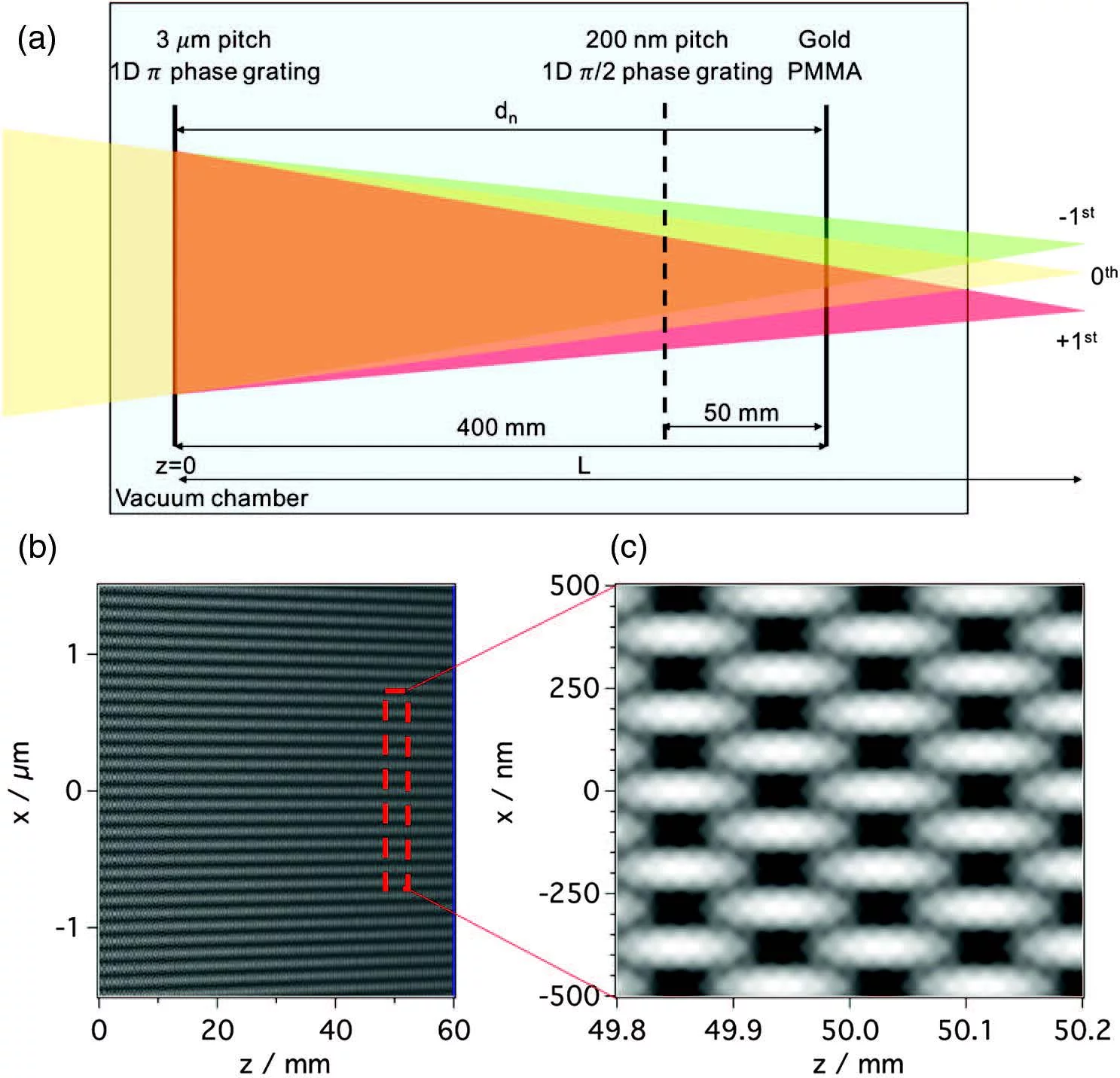
The extension of transient grating spectroscopy to the X-ray regime is very appealing, opening possibilities ranging from the study of thermal transport in the ballistic regime to charge, spin, and energy transfer processes with atomic spatial and femtosecond temporal resolution. Studies involving complicated split-and-delay lines have not yet been successful in achieving this goal. In an experiment at SwissFEL, X-ray transient gratings were prepared using a simple method based on the Talbot effect for converging beams. By analyzing printed interference patterns on polymethyl methacrylate and gold samples using ∼3 keV X-ray pulses, a the experimental feasibility transient gratings in the hard X-ray regime was demonstrated and published in Optics Letters.
TG excitation was demonstrated in the hard X-ray range at 7.1 keV. In bismuth germanate (BGO), the non-resonant TG excitation generates coherent optical phonons detected as a function of time by diffraction of an optical probe pulse. This experiment demonstrates the ability to probe bulk properties of materials and paves the way for ultrafast coherent four-wave-mixing techniques using X-ray probes and involving nanoscale TG spatial periods. The results are published in Nature Photonics.
Tackling the timing problem in ultrafast spectroscopy: Simultaneous single shot, time-resolved demagnetization dynamics at two energies
Streaking the time information in one dimension on a detector
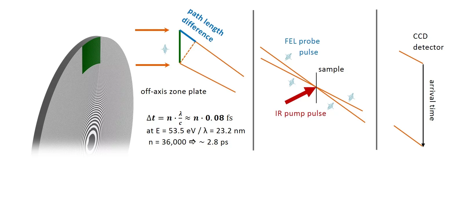
X-ray free electron lasers exhibit unique capabilities to conduct resonant x-ray spectroscopy techniques on ultrafast time scales. Utilizing diffractive optical elements, the pathway difference inherent to diffraction can be exploited to streak the arrival time of an x-ray probe along a geometric dimension. This concept has been applied in a pioneering experiment in reflection geometry at FLASH with a time resolution of 120 fs: Single-shot Monitoring of Ultrafast Processes via X-ray Streaking at a Free Electron Laser.
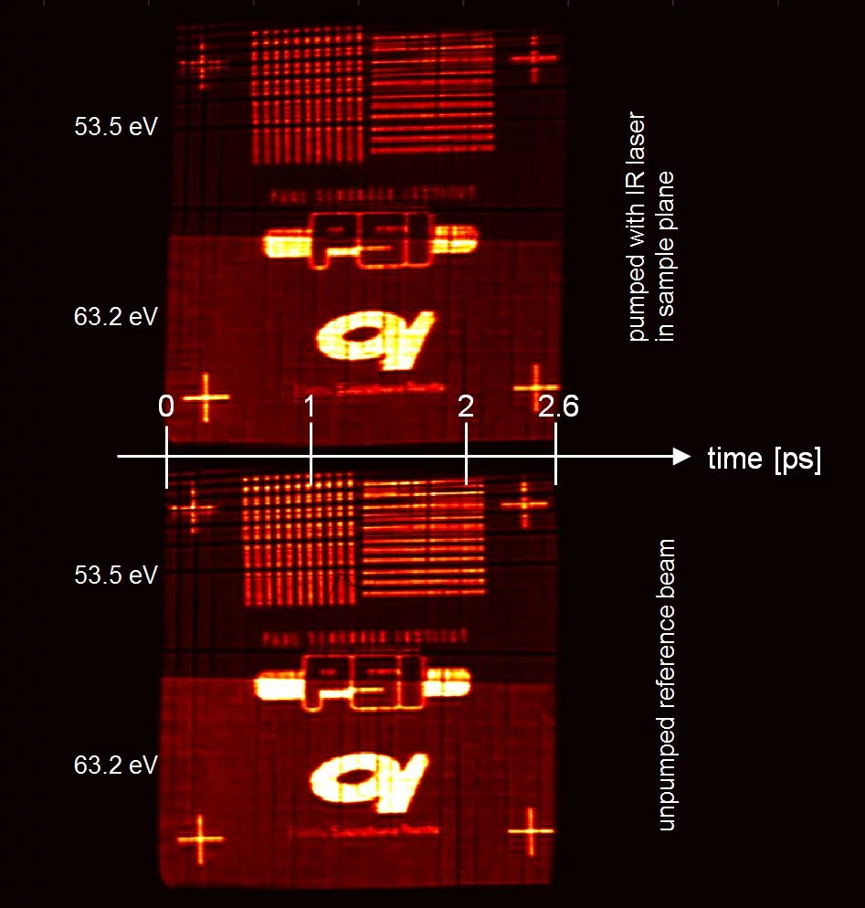
Adapting the experiment to a transmission geometry, we investigated demagnetization dynamics of a CoDy film together with collaborators from CNRS, the University Pierre and Marie Curie, and at the FERMI free electron laser, see figure above. The time window reachable with this setup is 1.5 ps, theoratically limited by the wavelenght divided through the speed of light. In practice, the duration of the pump puls of approx. 100 fs is the limiting factor.
Extending the concept to multiple energies
Extending this scheme to take advantage of the other spatial dimension, more advanced experiments become possible. In particular, we aim at performing ultrafast spectroscopy. Advancing towards this goal, we have designed a specific optical element, which allows to perform time-streaking experiments at two distinct energies simultaneously. This allows us to investigate multicomponent systems at two different absorption edges, and to compare dynamics of two elements with exactly the same timing (without a different time zero).
Employing this scheme, we investigated the demagnetization dynamics in iron-nickel multilayer systems and alloys. The evaluation of the data is ongoing. In principle, the number of different energies is not limited and can be extended even to a continuous energy range.
Selected Publications
For an extensive overview we kindly refer you to our publication repository DORA.
Hard X-ray helical dichroism of disordered molecular media
J. Rouxel, B. Rösner, D. Karpov, C. Bacellar, G. F. Mancini, F. Zinna, D. Kinschel, O. Cannelli, M. Oppermann, C. Svetina, A. Diaz, J. Lacour, C. David, M. Chergui, Nature Photonics 16, 2022, 570-574
https://doi.org/10.1038/s41566-022-01022-x
Chirality is a structural property of molecules lacking mirror symmetry that has strong implications in diverse fields, ranging from life sciences to materials science. Chirality-sensitive spectroscopic methods, such as circular dichroism, exhibit weak signal contributions on an achiral background. Helical dichroism, which is based on the orbital angular momentum (OAM) of light, offers a new approach to probe molecular chirality, but it has never been demonstrated on disordered samples. Furthermore, in the optical domain the challenge lies in the need to transfer the OAM of the photon to an electron that is localized on an ångström-size orbital. Here we overcome this challenge using hard X-rays with spiral Fresnel zone plates, which can induce an OAM. We present the helical dichroism spectra of a disordered powder sample of enantiopure salts of the molecular complex of [Fe(4,4′-diMebpy)3]2+ at the iron K edge (7.1 keV) with OAM-carrying beams. The asymmetry ratios for the helical dichroism spectra are within one to five percent for OAM beams with topological charges of one and three. These results open a new window into the studies of molecular chirality and its interaction with the OAM of light.
Observation of Magnetic Helicoidal Dichroism with Extreme Ultraviolet Light Vortices
Mauro Fanciulli, Matteo Pancaldi, Emanuele Pedersoli, Mekha Vimal, David Bresteau, Martin Luttmann, Dario De Angelis, Primož Rebernik Ribič, Benedikt Rösner, Christian David, Carlo Spezzani, Michele Manfredda, Ricardo Sousa, Ioan-Lucian Prejbeanu, Laurent Vila, Bernard Dieny, Giovanni De Ninno, Flavio Capotondi, Maurizio Sacchi, and Thierry Ruchon, Physical Review Letters 128, 2022, 077401
https://doi.org/10.1103/PhysRevLett.128.077401
CWe report on the experimental evidence of magnetic helicoidal dichroism, observed in the interaction of an extreme ultraviolet vortex beam carrying orbital angular momentum with a magnetic vortex. Numerical simulations based on classical electromagnetic theory show that this dichroism is based on the interference of light modes with different orbital angular momenta, which are populated after the interaction between light and the magnetic topology. This observation gives insight into the interplay between orbital angular momentum and magnetism and sets the framework for the development of new analytical tools to investigate ultrafast magnetization dynamics.
Light-Induced Magnetization at the Nanoscale
Jonas Wätzel, Primož Rebernik Ribič, Marcello Coreno, Miltcho B. Danailov, Christian David, Alexander Demidovich, Michele Di Fraia, Luca Giannessi, Klavs Hansen, Špela Krušič, Michele Manfredda, Michael Meyer, Andrej Mihelič, Najmeh Mirian, Oksana Plekan, Barbara Ressel, Benedikt Rösner, Alberto Simoncig, Simone Spampinati, Matija Stupar, Matjaž Žitnik, Marco Zangrando, Carlo Callegari, Jamal Berakdar, and Giovanni De Ninno, Physical Review Letters 128, 2022, 157205
https://doi.org/10.1103/PhysRevLett.128.157205
Triggering and switching magnetic moments is of key importance for applications ranging from spintronics to quantum information. A noninvasive ultrafast control at the nanoscale is, however, an open challenge. Here, we propose a novel laser-based scheme for generating atomic-scale charge current loops within femtoseconds. The associated orbital magnetic moments remain ferromagnetically aligned after the laser pulses have ceased and are localized within an area that is tunable via laser parameters and can be chosen to be well below the diffraction limit of the driving laser field. The scheme relies on tuning the phase, polarization, and intensities of two copropagating Gaussian and vortex laser pulses, allowing us to control the spatial extent, direction, and strength of the atomic-scale charge current loops induced in the irradiated sample upon photon absorption. In the experiment we used He atoms driven by an ultraviolet and infrared vortex-beam laser pulses to generate current-carrying Rydberg states and test for the generated magnetic moments via dichroic effects in photoemission. Ab initio quantum dynamic simulations and analysis confirm the proposed scenario and provide a quantitative estimate of the generated local moments.
Photoelectric effect with a twist
G. de Ninno, J. Wätzel, P. R. Ribič, E. Allaria, M. Coreno, M. B. Danailov, C. David, A. Demidovich, M. Di Fraia, L. Giannessi, K. Hansen, Š. Krušič, M. Manfredda, M. Meyer, A. Mihelič, N. Mirian, O. Plekan, B. Ressel, B. Rösner, A. Simoncig, S. Spampinati, M. Stupar, M. Žitnik, M. Zangrando, C. Callegari, J. Berakdar, Nature Photonics 14, 2020, 554-558
https://doi.org/10.1038/s41566-020-0669-y
Photons have fixed spin and unbounded orbital angular momentum (OAM). While the former is manifested in the polarization of light, the latter corresponds to the spatial phase distribution of its wave front. The distinctive way in which the photon spin dictates the electron motion upon light-matter interaction is the basis for numerous well-established spectroscopies that reveal the electronic, magnetic and structural properties of matter. In contrast, imprinting OAM on a matter wave, specifically on a propagating electron, is generally considered very challenging and the anticipated effect undetectable. Surprisingly, we find experimentally, and confirm theoretically, that the OAM of an optical field can be imprinted coherently onto a propagating electron wave, and that this phase information survives ensemble averaging out to macroscopic distances, where the electron is detected. We also show that electronic transitions, which are otherwise optically inaccessible due to selection rules, are essential for this process to occur. Our results reveal new aspects of light-matter interaction and point to a new kind of single-photon electron spectroscopy for accessing electronic optical transitions that are usually forbidden by symmetry.
7 nm spatial resolution in soft X-ray microscopy and high resolution magnetic imaging
B. Rösner, S. Finizio, F. Koch, F. Döring, V. A. Guzenko, A. Kleibert, M. Langer, E. Kirk, M. Meyer, J. L. Ornelas, A. Späth, R. H. Fink, S. Stanescu, S. Swaraj, R. Belkhou, B. Watts, J. Raabe, C. David, Optica 7, 2020, 1602-1608
https://doi.org/10.1364/OPTICA.399885
The availability of intense soft X-ray beams with tunable energy and polarization has pushed the development of highly sensitive, element-specific and non-invasive microscopy techniques to investigate condensed matter with high spatial and temporal resolution. The short wavelengths of soft X‑rays promise to reach spatial resolutions in the deep single-digit nanometer regime, providing unprecedented access to magnetic phenomena at fundamental length scales. Despite considerable efforts in soft X‑ray microscopy techniques, a two-dimensional resolution of ten nanometers has not yet been surpassed in direct imaging. Here, we report on a significant step beyond this long-standing limit by combining newly developed soft X-ray Fresnel zone plate lenses with advanced precision in scanning control and careful optical design. With this approach, we achieve an image resolution of seven nanometers. By combining this highly precise microscopy technique with the X-ray magnetic circular dichroism effect, we reveal dimensionality effects in an ensemble of interacting magnetic nanoparticles. Such effects are topical in current nanomagnetism research and highlight the opportunities of high resolution soft X-ray microscopy in magnetism research and beyond.


