Neutron imaging is a non-destructive testing (NDT) method allowing investigations in a variety of topics in the area of wood research. Of special importance in this field of research is the high sensitivity of neutron radiation for hydrogen. Even though hydrogen represents only a portion of ca. 6%wt in the elemental composition of wood it accounts for more than 90% of the neutron attenuation. The hydrogen content entails the high attenuation coefficients of wood, which account for the two beamlines: NEUTRA: 1.13cm-1 and ICON: 1.42cm-1; these values are theoretical attenuation coefficients for a simplified physical model of wood with ρdry = 0.6 g/cm3 (cf. table 1; source: [1]). Hence, wood shows good contrasts even for relatively small samples, but it also limits the potential size of wood samples to small and medium sized specimens of few centimetres. In its working range, neutron imaging yields results equivalent to X-ray allowing for example the analyses of density variations in annual growth rings [2, 3]. The highest spatial resolution available up to the present for neutron imaging (pixel size 13.5µm/px) is nevertheless not yet sufficient to resolve the wood structure on a cellular level with exception of large (earlywood) vessels.
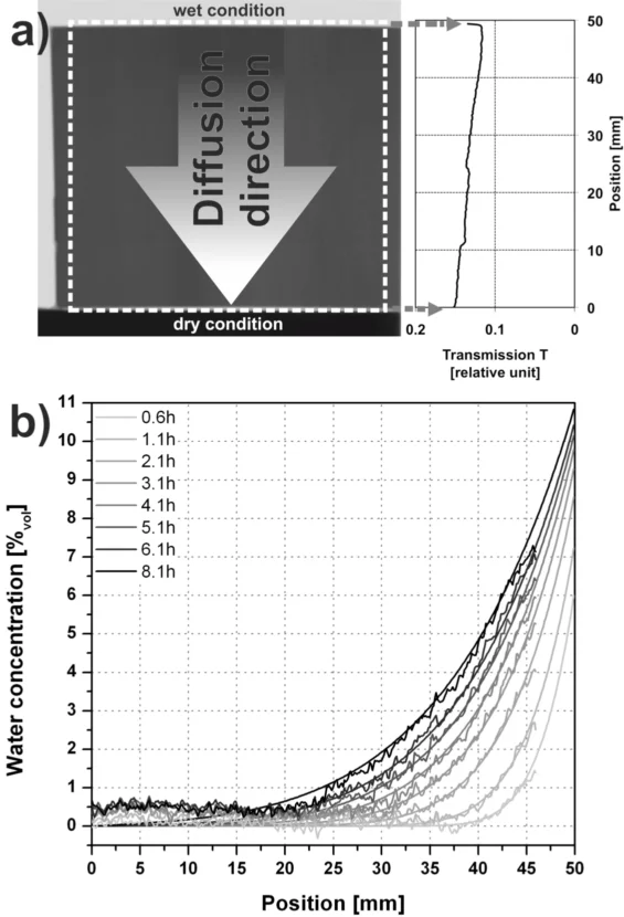
Due to the high sensitivity for hydrogen, neutron imaging is also particularly suited for investigations on the moisture content within wood, which is of special interest as most of the wood properties are directly or indirectly related to it. Neutron imaging allows the dynamic assessment of moisture transport processes, both qualitatively as well as quantitatively [4, 5]. The high attenuation coefficient of water allows to detect water layers with a total thickness (summed up along the beam path through the sample) of 40 to 50 µm (depending on the neutron spectrum), which correspond to moisture content changes (Δu) of ca. 0.9% for NEUTRA and 0.7% for ICON for wood samples with 1 cm thickness in beam direction and a dry density of 0.6 g/cm3. Similar results can be expected for dynamic investigations on the interaction of wood with other hydrogenous materials such as adhesives, lacquers, varnishes, etc. A detailed overview on the possibilities and limitations of neutron imaging with concern of wood research is given in [6].
Publications
[1] Neutron attenuation coefficients for non-invasive quantification of wood properties.
Mannes, D., Josic, L., Lehmann, E. & Niemz, P. (2009)
Holzforschung, 63, 472-478.
DOI: 10.1515/hf.2009.081
[2] Neutron imaging versus standard X-ray densitometry as method to measure tree-ring wood density.
Mannes, D., Lehmann, E., Cherubini, P. & Niemz, P. (2007)
Trees-Structure and Function, 21, 605-612.
DOI: 10.1007/S00468-007-0149-8
[3] Silviscan Vs. Neutron Imagin to Generate Radial Softwood Density Profiles.
Keunecke, D., Mannes, D., Niemz, P., Lehmann, E. & Evans, R. (2010)
Wood Research, 55, 49-59.
[4] Non-destructive determination and quantification of diffusion processes in wood by means of neutron imaging.
Mannes, D., Sonderegger, W., Hering, S., Lehmann, E. & Niemz, P. (2009)
Holzforschung, 63, 589-596.
DOI: 10.1515/Hf.2009.100
[5] Quantitative determination of bound water diffusion in multilayer boards by means of neutron imaging.
Sonderegger W, Hering S, Mannes D, Vontobel P, Lehmann E, Niemz, (2010)
EUROPEAN JOURNAL OF WOOD AND WOOD PRODUCTS 68, 341 (2010)., 68, 341.
DOI: 10.1007/s00107-010-0463-5
[6] Non-destructive testing of wood by means of neutron imaging in comparison with similar methods.
Mannes, D.
PhD Thesis ETH (2009).
DOI: 10.3929/ethz-a-005916121


![Figure 1: Section of a partially degraded soft-wood sample in a neutron transmission image. The 500-year-old sample originates from an excavation near Dachstein (Austria) [6].](/sites/default/files/styles/primer_slick_scale/public/import/niag/WoodEN/Figure1.png.webp?itok=I3JL7bri)
![Figure 2: Tomography of an oak wood sample (Quercus spp.) using the µ-CT-setup at ICON; left side: reconstructed axial slice through showing perspicuous differences between the tissues (water conducting areas, fibre tissue and wood rays); right side: visualised CT-data showing the 3D-distribution of the different tissues [6].](/sites/default/files/styles/primer_slick_scale/public/import/niag/WoodEN/Figure2.png.webp?itok=ic6UlubA)

![Figure 4: Neutron tomogram through specimens of beech wood (left) and spruce wood (right) with a 1K-PUR adhesive joint; the glue line appears pronounced due to the high sensitivity for hydrogen [6].](/sites/default/files/styles/primer_slick_scale/public/import/niag/WoodEN/Figure4.png.webp?itok=GTAxXhVo)