The first prototype of PSI's neutron microscope utilizes a high-NA magnifying optics and about 4 μm thin scintillator based on nat-Gd2O2S:Tb. The images of gadolinium Siemens star test object show spatial resolution of 7.6 μm – an improvement of a factor 4 over the spatial resolution available by standard high-resolution imaging using hitherto available at PSI [1, 2]. Further details are available in Trtik et al. [1].
Isotopically enriched 157-Gd2O2S:Tb scintillator screens were developed within the framework of the neutron microscope project. For about 2.5 μm thickness of the scintillator screens, the enrichment leads to the improvement in the absorption and the light output by factor 3.8 and 3.6, respectively. Further details available in Trtik et al. [3].
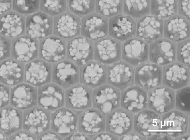
We are currently working on various aspect of the project (e.g. development of microstructured neutron-sensitive scintillators).
Publications
[1] Improving the spatial resolution of neutron imaging at Paul Scherrer Institut – The Neutron Microscope Project
Pavel Trtik, Jan Hovind, Christian Gruenzweig, Alex Bollhalder, Vincent Thominet, Christian David, Anders Kaestner, and Eberhard H. Lehmann.
Physics Procedia, 69, 169 (2015)
DOI: 10.1016/j.phpro.2015.07.024
[2] The ICON beamline - A facility for cold neutron imaging at SINQ
Kaestner, AP; Hartmann, S; Kuehne, G; Frei, G; Gruenzweig, C; Josic, L; Schmid, F ; Lehmann, EH
NUCLEAR INSTRUMENTS & METHODS IN PHYSICS RESEARCH SECTION A, 659 (2011), p. 387-393
DOI: 10.1016/j.nima.2011.08.022
[3] Isotopically-enriched gadolinium-157 oxysulfide scintillator screens for the high-resolution neutron imaging
Trtik, P., Lehmann, E. H.
NUCLEAR INSTRUMENTS & METHODS IN PHYSICS RESEARCH SECTION A, 788 (2015), p. 67-70
DOI: 10.1016/j.nima.2015.03.076

![Comparison of the images of the gadolinium Siemens star test object using (left top) the standard MICRO-setup, (right) the neutron microscope, and (left bottom) an electron microscope . For details, please see Trtik et al [1].](/sites/default/files/styles/primer_full_image_xxl/public/import/niag/NeutronMicroscopeEN/Figure1.jpg.webp?itok=xUE0NrFF)
![Comparison of the light output and the absorption of the isotopically enriched (Gd-157) and the natural Gd2O2S:Tb scintillator screens. For details, please see Trtik et al [3].](/sites/default/files/styles/primer_full_image_xxl/public/import/niag/NeutronMicroscopeEN/Figure2.jpg.webp?itok=LIORZTOE)
