• Paul Scherrer Institut PSI
  • PSI Research, Labs & User Services

Digital User Office

  • Digital User Office
  • DE
  • EN
  • FR
Paul Scherrer Institut (PSI)
Search
Paul Scherrer Institut (PSI)

Hauptnavigation

  • Research at PSIOpen mainmenu item
    • Research Initiatives
    • Ethics and Research integrity
    • Scientific Highlights
    • Scientific Events
    • Scientific Career
    • PSI-FELLOW
    • PSI Data Policy
  • Research Divisions and LabsOpen mainmenu item
    • Overview
    • Research with Neutrons and Muons
    • Photon Science
    • Energy and Environment
    • Nuclear Energy and Safety
    • Biology and Chemistry
    • Scientific Computing, Theory and Data
    • Large Research Facilities
  • Facilities and InstrumentsOpen mainmenu item
    • Overview
    • Large Research Facilities
    • Facilities
    • PSI Facility Newsletter
  • PSI User ServicesOpen mainmenu item
    • User Office
    • Methods at the PSI User Facilities
    • Proposals for beam time
    • Proposal Deadlines
    • Data Analysis Service (PSD)
    • EU support programmes
  • New ProjectsOpen mainmenu item
    • SLS 2.0
    • IMPACT
  • DE
  • EN
  • FR

Digital User Office (mobile)

  • Digital User Office

You are here:

  1. PSI Home
  2. Labs & User Services
  3. NUM
  4. LNS
  5. NIAG
  6. What is Neutron Imaging ?
  7. Neutron Physics
  8. Comparison to X-ray

Secondary navigation

Neutron Imaging and Applied Materials

  • People
  • What is Neutron Imaging ? Expanded submenu item
    • Neutron Imaging Setup
    • Neutron Physics Expanded submenu item
      • Neutron Interaction with Matter
      • Comparison to X-ray
    • Neutron Imaging Techniques and Methods
      • Neutron Imaging Detectors
      • Neutron Tomography
      • Dynamic Neutron Imaging
      • Advanced Neutron Imaging Techniques
  • Beamlines
  • Research and Development
  • Industrial Services
  • Gallery
    • Movies
    • Bronze Sculptures
  • Publications
  • News / Events
    • News Archive
  • Links

Comparison to X-ray

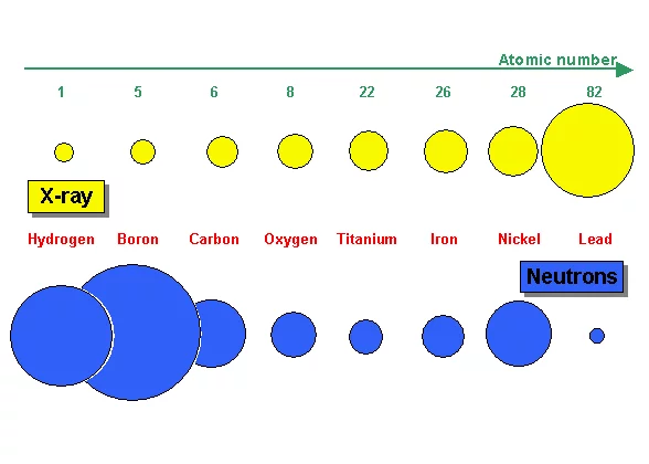
Figure 7 a: Comparison of the attenuation characteristics of selected materials for neutrons and X-ray
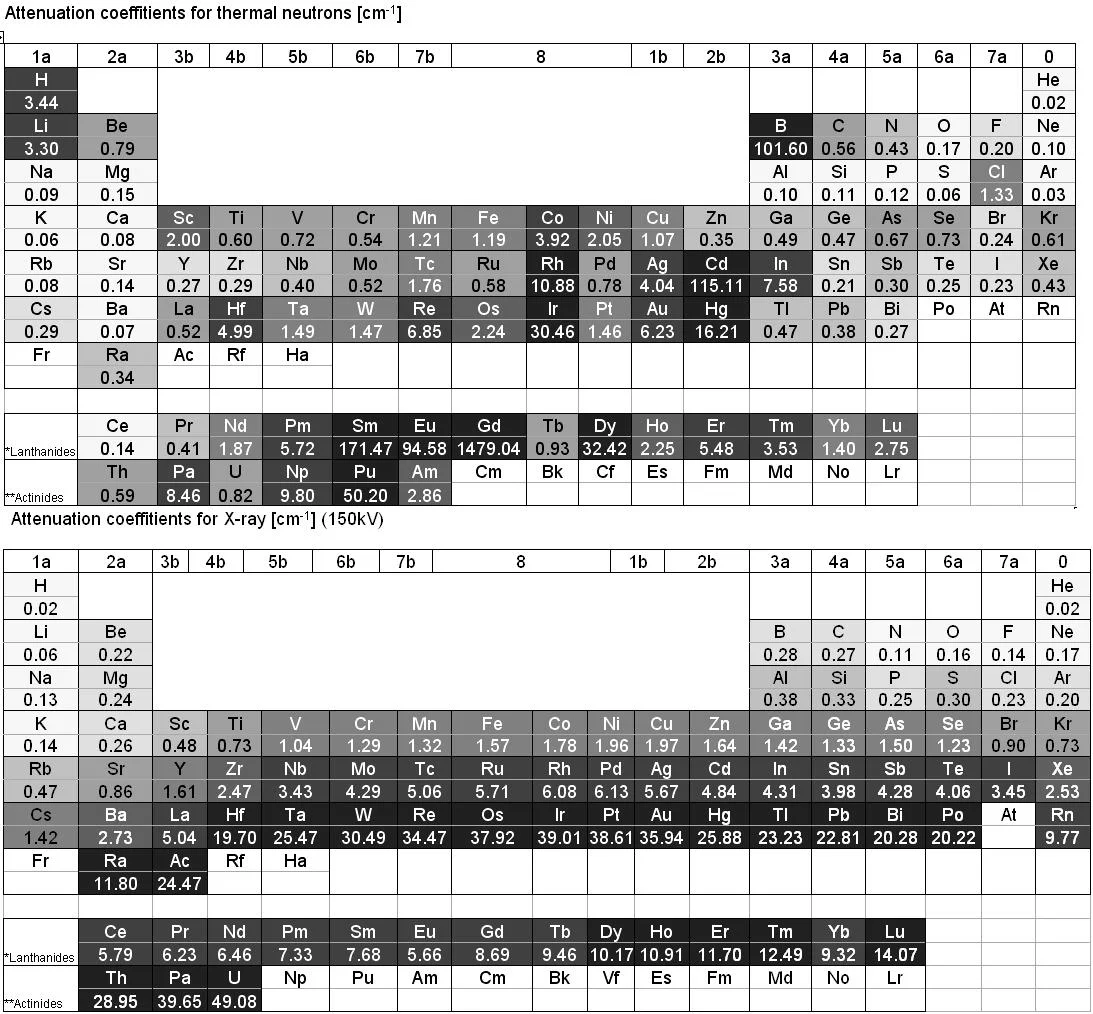
Figure 7 b: Comparison of the linear attenuation coeffitients for thermal neutrons (top) to 150 keV X-ray (bottom)
X-ray is electromagnetic radiation which mainly interacts with the electrons of an atom. Thus the more electrons an atom has (i.e. with increasing atomic number Z) the higher the probability for X-ray interaction as shown in Figure 7a. X-ray is therefore an excellent imaging modality for biolological material e.g the human body containing a lot of water. Quite contrary, neutron interaction probability is high in water due to the high scattering probability with the hydrogen atom. Thus a 1 cm thick layer of water is almost opaque for thermal neutrons. The neutron as an electrically neutral particle rather interacts with the atomic nucleus. It is able to transmit thick layers e.g. of metallic material as shown e.g. for lead in Figure 7a and b.

The different radiation matter interaction characteristic is the reason for another effect i.e. activation of nuclei by neutron capture producing instable (mainly short lived) radioactive isotopes. Samples have to be tested for radioactivity after neutron imaging before they can leave the beamline.

For imaging the quite different type of X-ray and neutron source characteristics induce further discrepancies. Due to very high brightness of e.g. synchrotron radiation sources very high spatial resolutions are possible i.e. below 1 µm. For µ-focus X-ray tubes enlarging cone-beam optics are feasible, which allows to get high spatial resolution for small samples. Neutron sources have comparably lower source brightness and extracted neutron beams cannot easily be focussed down to spot sizes below 1 mm. Neutron imaging therefore relies mainly on parallel beam irradiation and effective spatial resolution is often limited by the thickness of the scintillator (some 10 µm's as thinnest layer).

Sidebar

Neutron Imaging Brochure

NI Brochure
Read more


Download Flyer

Contact

Applied Materials Group
Paul Scherrer Institut
LNS WBBA/118
5232 Villigen PSI

Prof. Dr. Markus Strobl
Group leader

Phone: +41 56 310 5941
E-mail: markus.strobl@psi.ch

Secretary: R. Bercher
Phone: +41 56 310 3402
Fax: +41 56 310 3131
E-mail: renate.bercher@psi.ch
 

Additional Information

  • Research with neutrons, synchrotron light and muons
    More about the most important tools in PSI's research on the structure of matter.
  • Research with PSI's large-scale facilities
    The institute's main experimental facilities
top

Footer

Paul Scherrer Institut

Forschungsstrasse 111
5232 Villigen PSI
Switzerland

Telephone: +41 56 310 21 11
Telefax: +41 56 310 21 99

How to find us
Contact

Visitor Centre psi forum
School Lab iLab (in German)
Center for Proton Therapy
PSI Education Centre
PSI Guest House
PSI Gastronomie (in German)
psi forum shop

Service & Support

  • Phone Book
  • User Office
  • Accelerator Status
  • PSI Publications
  • Suppliers
  • E-Billing
  • Computing
  • Safety (in German)

Career

  • Working at PSI
  • Job Opportunities
  • Training and further education
  • Career Center
  • Vocational Training (in German)
  • PSI Education Center

For the media

  • PSI in brief
  • Facts and Figures
  • Media corner
  • Media Releases
  • Social Media

Follow us: Twitter (in English) LinkedIn Youtube Facebook Instagram Issuu RSS

Footer legal

  • Imprint
  • Terms and Conditions
  • Editors' login