
Scientific Highlights
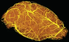
Hierarchical imaging and computational analysis of three-dimensional vascular network architecture in mouse brain
An international team involving researchers from the University and University Hospital Zürich, the Krembil Research Institute and the University and University Hospital in Toronto (Canada), the Department of Physics of Jyväskylä (Finland), the University of Leuven (Belgium), the Johannes Kepler University in Linz (Austria), the Novartis Institutes for Biomedical Research in Emeryville (USA), the ETH Zürich and the Paul Scherrer Institute has developed a protocol that enables hierarchical imaging and computational analysis of vascular networks in entire postnatal- and adult mouse brains, enabling direct and quantitative comparisons of the morphological brain vascular network architecture between different postnatal and / or adult developmental stages. The results have been published on Nature Protocols on September 3rd, 2021.
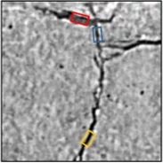
Understanding Why Solid-State Batteries Fail
Researchers from the University of Oxford, the Diamond Light Source and the Paul Scherrer Institut have generated strong evidence supporting one of two competing theories regarding the mechanism by which lithium metal dendrites grow through ceramic electrolytes. A process leading to short circuit at high rates of charge. The X-ray phase-contrast imaging capabilities of the TOMCAT beamline of the Swiss light source allowed researchers to visualize and characterize the growth of cracks and dendrites deep within an operating solid-state battery. The results were published in Nature Materials on April 22, 2021.
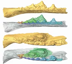
Deep evolutionary origins of the human smile
Detailed characterization of the tooth and jaw structure and development among shark ancestors by synchrotron based X-ray tomographic microscopy at TOMCAT led an international team of researchers from the Naturalis Biodiversity Center in Leiden and the University of Bristol to the discovery that while teeth evolved once, complex dentitions have been gained and lost many times in evolutionary history.

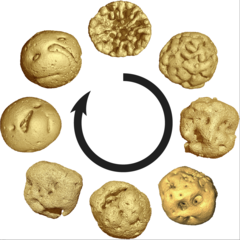
3D printing silica aerogels at the micrometer scale
A group of EMPA and ETH Zürich researchers have developed a new method to directly write ink made of silica aerogels in 3D. Thanks to X-ray phase contrast tomography at the TOMCAT beamline they characterized the resulting printed material with different compositions. Their results were published in Nature on August 18, 2020.
")
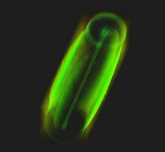
Phase contrast microtomography reveals nanoparticle accumulation in zebrafish
Metal-based nanoparticles are a promising tool in medicine – as a contrast agent, transporter of active substances, or to thermally kill tumor cells. Up to now, it has been hardly possible to study their distribution inside an organism. Researchers at the University of Basel in collaboration with the TOMCAT team have used phase contrast X-ray tomographic microscopy to take high-resolution captures of the nanoparticle aggregation inside zebrafish embryos.
The study was published in the journal Small and featured on the cover of its current issue.
Miniaturized fluidic circuitry observed in 3D
The team of Prof. Thomas Hermans at the University of Strasbourg in France managed to create wall-less aqueous liquid channels called anti-tubes. Thanks to X-ray phase contrast tomography at the TOMCAT beamline those anti-tubes could be observed in 3D. The exciting results were published in Nature on May 6, 2020.

X-ray Imaging for Biomedicine: Imaging Large Volumes of Fresh Tissue at High Resolution
The TOMCAT beamline at the Swiss Light Source specializes in rapid high-resolution 3-dimensional tomographic microscopy measurements with a strong focus on biomedical imaging. The team has recently developed a technique to acquire micrometer-scale resolution datasets on the entire lung structure of a juvenile rat in its fresh natural state within the animal’s body and without the need for any fixation, staining or other alteration that would affect the observed structure (E. Borisova et al., 2020, Histochem Cell Biol).

MacEtch in gas phase: a new nanofabrication technology at PSI
The grating fabrication team of the X-ray tomography group has scored another record in etching technology of silicon by realizing a MacEtch process in gas phase. Ultra-high aspect ratios (up to 10 000 : 1) in the nanoscale regime (down to 10 nm) were achieved by platinum assisted chemical etching of silicon in the gas phase. The results were published in Nanoscale Horizons on February 17, 2020.
Animal embryos evolved before animals
Detailed characterization of cellular structure and development of exceptionally preserved ancient tiny fossils from South China by synchrotron based X-ray tomographic microscopy at TOMCAT led an international team of researchers from the University of Bristol and Nanjing Institute of Geology and Palaeontology to the discovery that animal-like embryos evolved long before the first animals appear in the fossil record.