Show filters
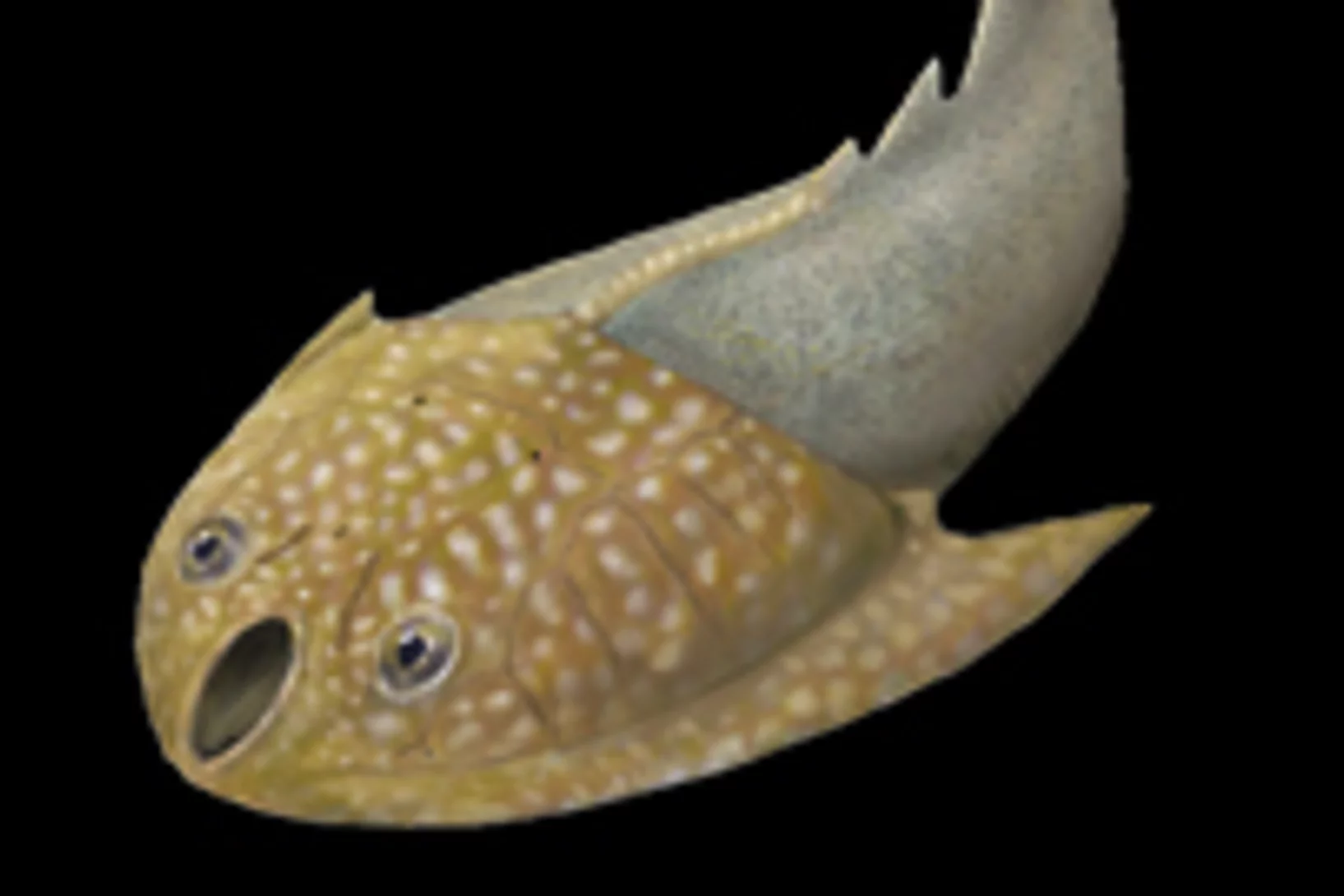
Unseren frühen Vorfahren in den Kopf (und in die Nase) geschaut
Der Umbau des Gehirns und der Sinnesorgane dürfte den Erfolg der Wirbeltiere, eines der grossen Rätsel der Evolutionsbiologie, erklären à so die Aussage einer Arbeit, die heute im Wissenschaftsjournal Nature erschienen ist. Die Forschenden konnten das Rätsel durch Untersuchungen des Gehirns eines 400 Millionen Jahre alten versteinerten Fisches à eines evolutionären Bindeglieds zwischen den heute lebenden kiefertragenden Wirbeltieren und den Kieferlosen.
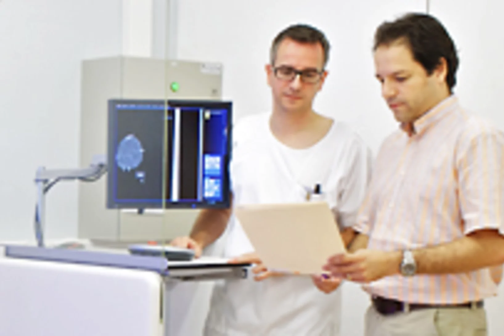
Neue Methode für die Krebserkennung mit Brustgewebe erprobt
Das Paul Scherrer Institut PSI hat eine neue Methode zur Diagnose von Brustkrebs entwickelt und nun zusammen mit dem Kantonsspital Baden AG erstmals an nicht-konserviertem, menschlichem Gewebe erprobt. Dabei wurde erkannt, dass es mit der neuen Methode möglich sein sollte, Strukturen sichtbar zu machen, die mit der herkömmlichen Mammografie nicht abgebildet werden. Wissenschaftler der Forschungsabteilung des Unternehmens Philips untersuchen derzeit auf Grundlage des vorgestellten Verfahrens den Einsatz in der medizinischen Praxis.
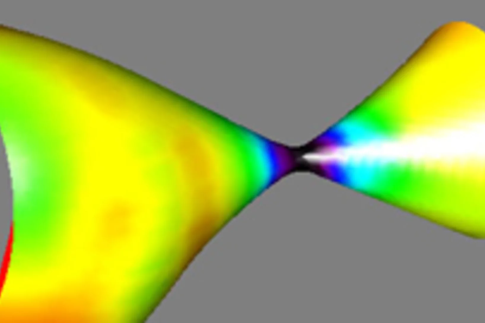
Universelles Gesetz für Veränderungen in Werkstoffen gefunden
In vielen wichtigen Werkstoffen findet man mehrere Phasen. Wird ein solcher Werkstoff erwärmt, können Atome von der einen Phase zur anderen wandern, so dass sich die Verteilung der Phasen ändert à und damit oft die Eigenschaften des Werkstoffs. Nun haben Forschende für einen wichtigen Fall einer solchen Veränderung gezeigt, dass es eine universelle Gesetzmässigkeit gibt, die den Vorgang beschreibt. Und zwar für alle Werkstoffklassen.
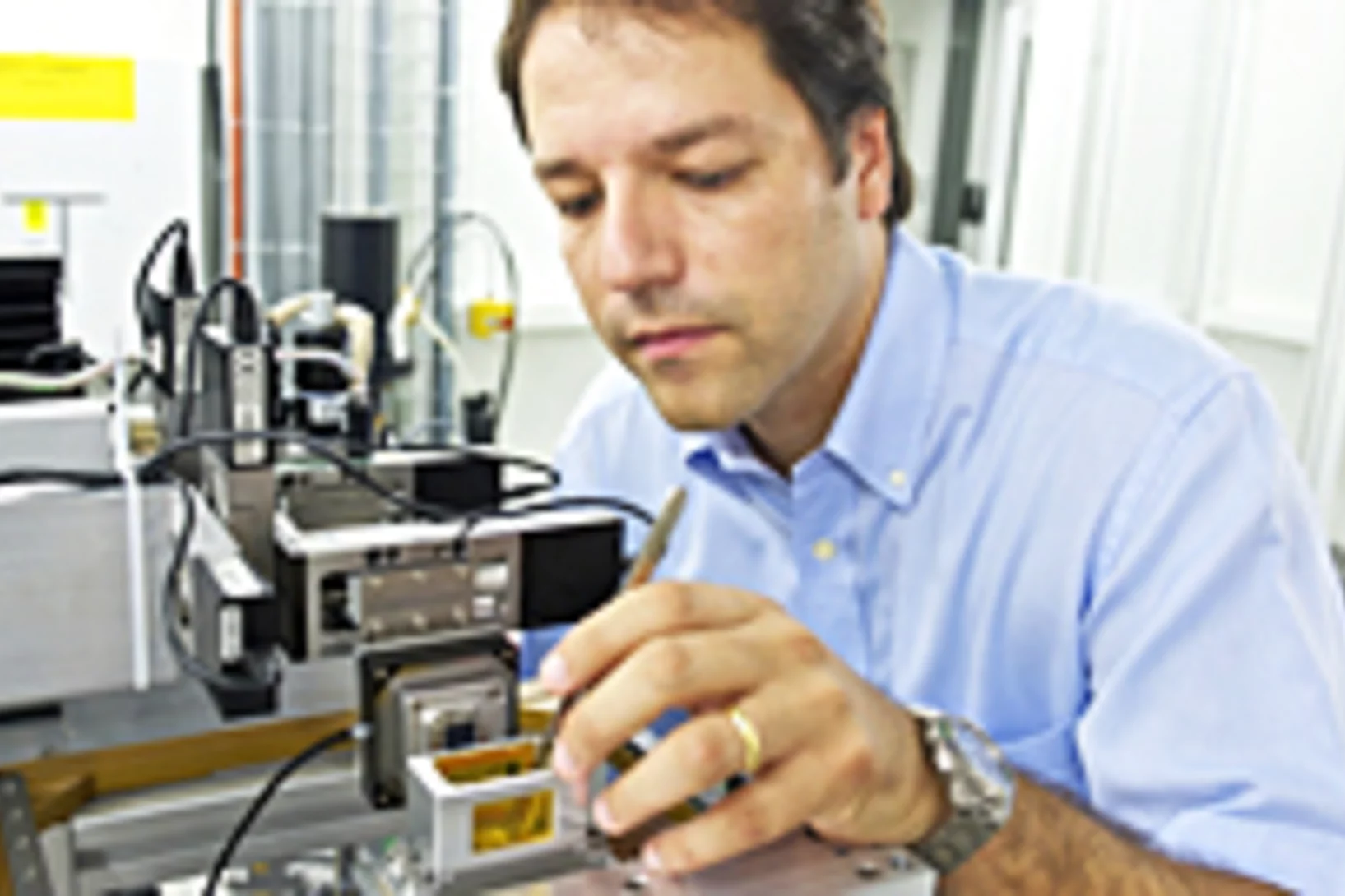
Neues Röntgenverfahren unterscheidet, was bisher gleich aussah
Auf Bildern, die mit Phasenkontrastverfahren erzeugt werden, kann man Gewebe unterscheiden, das auf gewöhnlichen Röntgenbildern fast gleich aussieht: etwa Muskeln, Knorpel, Sehnen oder Weichteiltumore. Forschende des Paul Scherrer Instituts und der Chinesischen Akademie der Wissenschaften haben das Verfahren so weiterentwickelt, dass es in Zukunft einfacher zu handhaben sein wird. Das könnte helfen, Tumore zu erkennen oder gefährliche Gegenstände im Gepäck sichtbar zu machen.