
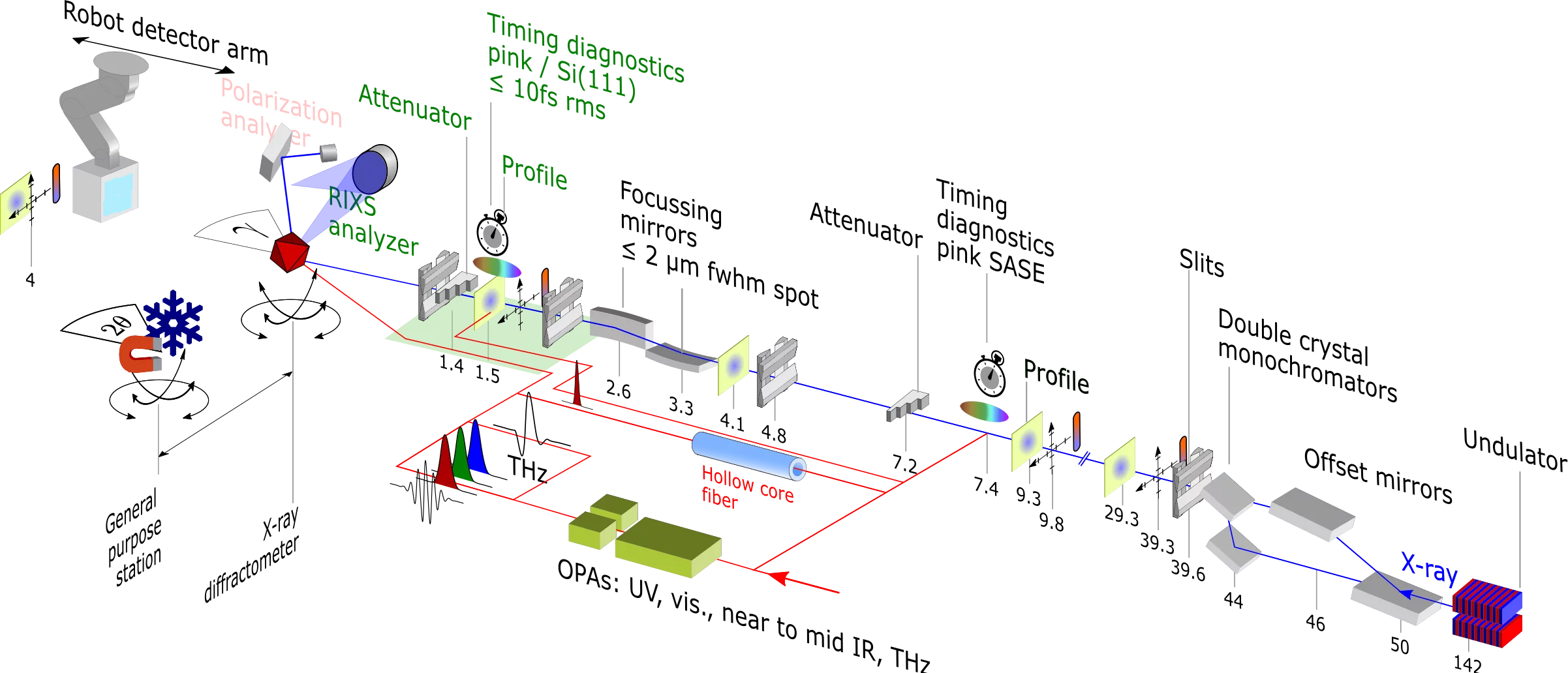
Schematic Bernina Instrument at the center SwissFEL Aramis beamline.
The Bernina instrument is formed by the center SwissFEL Aramis beamline, the pump laser system, and different endstations.
Besides versatile setups using the modular diffractometer system, the standardized setups
can be used for experiments.
| Bernina endstation and X-ray parameters | |||
|---|---|---|---|
| Photon energy range | 2 keV – 13.0 keV | ||
| Beam profile | Focused down to 2x2 µm2 (fwhm, measured) to unfocused 1000x1000 µm2 (fwhm, photon energy dependent). | ||
| Bandwidth | Monochromatic (Si(111) routinely used, InSb(111), Si(311) and pink beam (~0.2% of fundamental, transmissive single FEL pulse spectrometer available), special modes like broadband SASE operation possible. | ||
| Pulse length | Standard SASE pulse length ~50 fs (fwhm), short pulse options at cost of pulse energy down to ~20 fs. | ||
| Environment | He or ambient atmosphere, platform for user-supplied chambers, N2 and He based cryostream coolers down to ~80 K. Vacuum chamber available for low sample Temperature (<5 K), high field THz excitation, and tender X-ray range. | ||
| Sample systems | Solids: single crystals, powders, amorphous systems. Liquid/Gas only with user supplied equipment. | ||
| Detectors and Spectrometer |
| ||
Beamline layout
Following table lists distances of significant beamline components from the sample location.
| Beamline component | Position along Bernina beamline (swissfel z direction) | |
relative to straight beam sample interaction position | relative to standard KB deflection sample interaction position (theta = 4 mRad) | |
| Profile monitor downstream of sample. | 3725 mm | 3716 mm |
| Straight beam sample location | 0 | -9.18 |
| Sample location at 4mRad KB mirror angles (standard) | 9.18 mm | 0 |
| Diamond window at Upstream diagnostics exit | -1330 mm |
|
| Attenuator targets in upstream diagnostics (att_usd) | -1420 mm |
|
| Target of time tool (tt_kb) / Profile monitor (prof_kb) | -1520 mm |
|
| Diamond window at Upstream diagnostics entry | -1945 mm |
|
| Horizontally deflecting/focusing KB mirror | -2600 mm |
|
| Vertically deflecting/focusing KB mirror | -3350 mm |
|