Direkt zum Inhalt
  • Paul Scherrer Institut PSI
  • PSI Research, Labs & User Services

Digital User Office

  • Digital User Office
  • DE
  • EN
  • FR
Paul Scherrer Institut (PSI)
Suche
Paul Scherrer Institut (PSI)

Hauptnavigation

  • Research at PSIÖffnen dieses Hauptmenu Punktes
    • Research Initiatives
    • Ethics and Research integrity
    • Scientific Highlights
    • Scientific Events
    • Scientific Career
    • PSI-FELLOW
    • PSI Data Policy
  • Research Divisions and LabsÖffnen dieses Hauptmenu Punktes
    • Overview
    • Research with Neutrons and Muons
    • Photon Science
    • Energy and Environment
    • Nuclear Energy and Safety
    • Biology and Chemistry
    • Scientific Computing, Theory and Data
    • Large Research Facilities
  • Facilities and InstrumentsÖffnen dieses Hauptmenu Punktes
    • Overview
    • Large Research Facilities
    • Facilities
    • PSI Facility Newsletter
  • PSI User ServicesÖffnen dieses Hauptmenu Punktes
    • User Office
    • Methods at the PSI User Facilities
    • Proposals for beam time
    • Proposal Deadlines
    • Data Analysis Service (PSD)
    • EU support programmes
  • New ProjectsÖffnen dieses Hauptmenu Punktes
    • SLS 2.0
    • IMPACT
  • DE
  • EN
  • FR

Digital User Office (mobile)

  • Digital User Office

Sie befinden sich hier:

  1. PSI Home
  2. Labs & User Services
  3. PSD
  4. SLS
  5. CSAXS
  6. Neurodegenerative disease studied by cryogenic X-ray nanotomography

Sekundäre Navigation

CSAXS

  • Status
  • User Information
    • Application for beamtime
    • Operation Schedule
    • Manuals
    • Software
    • Infrastructure
  • People
  • Beamline Layout
    • Source
    • Optics
  • Endstations
  • Research
  • Publications
  • Scientific Highlights
  • Contacts

Info message

Dieser Inhalt ist nicht auf Deutsch verfügbar.
4. November 2021

Neurodegenerative disease studied by cryogenic X-ray nanotomography

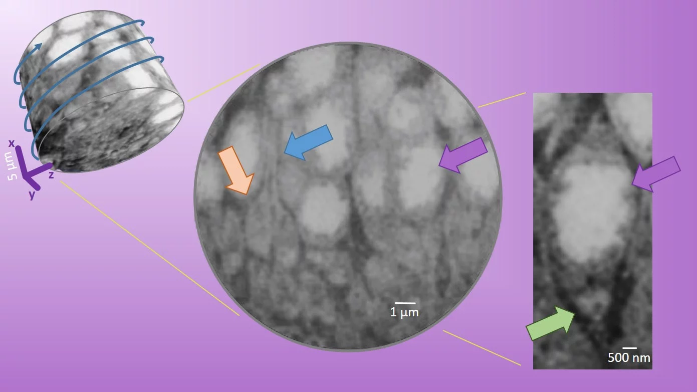
In a new study on retinal degeneration linked to blindness, a team of collaborators from the photon-science and biology divisions of PSI,  University of Zürich, and ETHZ, could see structures inside the nerve cells of the retina of a mouse. Biological tissue samples were imaged using ptychographic X-ray computer tomography (PXCT) at a resolution which allows to follow in three dimensions the axons path (blue arrow) of the retina in 3D. The technique allows to observe subcellular elements of the synapses (orange arrow) like the large mitochondria and the specialized ribbon responsible for neurotransmission, at an isotropic resolution of 150-200 nm.

Retina
Figure 1. Three views of a retina sample (plexiform layer of the outer retina) from wild type mouse shown at different magnifications. Left side: 3D rendering of a part of the sample showing the large nuclei of the photoreceptor cells with the high-density chromatin (white = high density). Many other smaller interlaced features corresponding to the plexiform layer are displayed. Middle: typical orthoslice showing nuclei (purple arrow) and the axonal fibers (blue arrow) terminated by the synaptical ends (orange arrow). Right side: zoom on a somatic synapse (green arrow) found in the cell body of a neuron.

Retinas from models of neurodegenerative disease were compared to the wild-type tissue. In the transgenic mouse model of retinitis pigmentosa, most synapses and axonal fibers of the photoreceptor cells are absent, and fibrosis prevails. The tomograms reveal the arrangement of more than 60 cell bodies in a cylindrical volume of 25 µm diameter and 20 µm height (upper left in figure 1). Because PXCT is a non-destructive technique, the researchers could perform complementary analysis of the sample from a healthy mouse at higher resolution with sequential nanometer slicing using a focused-ion beam and imaging with a scanning electron microscope. X-ray nanotomography  showed a good match of the subcellular structures like the ribbon synapse and even a somatic synapse.

The study paves the way for correlative analysis of neural tissues subject to neurodegeneration, remodeling and blindness treatments.

The work is online here and is selected as Research Highlight: Non-destructive imaging of large tissue volumes using X-rays

Read the full story

Imaging of retina cellular and subcellular structures using ptychographic hard X-ray tomography.
Panneels V, Diaz A, Imsand C, Guizar-Sicairos M, Müller E, Bittermann AG, Ishikawa T, Menzel A, Kaech A, Holler M, Grimm C, Schertler G.
J Cell Sci. 2021 134(19):jcs258561. doi: 10.1242/jcs.258561. Epub 2021 Oct 14. PMID: 34494099

Contact

Dr. Ana Diaz,
Swiss Light Source
Paul Scherrer Institut, 5232 Villigen PSI, Switzerland
Phone: +41 56 310 5626, e-mail: ana.diaz@psi.ch
https://www.psi.ch/en/sls/csaxs

Dr. Valérie Panneels,
Laboratory of Biomolecular Research
Paul Scherrer Institut, 5232 Villigen PSI, Switzerland
Phone: +41 56 310 2104, e-mail: valerie.panneels@psi.ch
https://www.psi.ch/en/lbr

 

Mit Sidebar

top

Fussbereich

Paul Scherrer Institut

Forschungsstrasse 111
5232 Villigen PSI
Schweiz

Telefon: +41 56 310 21 11
Telefax: +41 56 310 21 99

Der Weg zu uns
Kontakt

Besucherzentrum psi forum
Schülerlabor iLab
Zentrum für Protonentherapie
PSI Bildungszentrum
PSI Guest House (in english)
PSI Gastronomie
psi forum-Shop


Service & Support

  • Telefonbuch
  • User Office
  • Accelerator Status
  • Publikationen des PSI
  • Lieferanten
  • E-Rechnung
  • Computing
  • Sicherheit

Karriere

  • Arbeiten am PSI
  • Stellenangebote
  • Aus- und Weiterbildung
  • Career Center
  • Berufsbildung
  • PSI Bildungszentrum

Für die Medien

  • Das PSI in Kürze
  • Zahlen und Fakten
  • Mediacorner
  • Medienmitteilungen
  • Social Media

Folgen Sie uns: Twitter (deutsch) LinkedIn Youtube Facebook Instagram Issuu RSS

Footer legal

  • Impressum
  • Nutzungsbedingungen
  • Editoren-Login