Direkt zum Inhalt
  • Paul Scherrer Institut PSI
  • PSI Research, Labs & User Services

Digital User Office

  • Digital User Office
  • DE
  • EN
  • FR
Paul Scherrer Institut (PSI)
Suche
Paul Scherrer Institut (PSI)

Hauptnavigation

  • Research at PSIÖffnen dieses Hauptmenu Punktes
    • Research Initiatives
    • Ethics and Research integrity
    • Scientific Highlights
    • Scientific Events
    • Scientific Career
    • PSI-FELLOW
    • PSI Data Policy
  • Research Divisions and LabsÖffnen dieses Hauptmenu Punktes
    • Overview
    • Research with Neutrons and Muons
    • Photon Science
    • Energy and Environment
    • Nuclear Energy and Safety
    • Biology and Chemistry
    • Scientific Computing, Theory and Data
    • Large Research Facilities
  • Facilities and InstrumentsÖffnen dieses Hauptmenu Punktes
    • Overview
    • Large Research Facilities
    • Facilities
    • PSI Facility Newsletter
  • PSI User ServicesÖffnen dieses Hauptmenu Punktes
    • User Office
    • Methods at the PSI User Facilities
    • Proposals for beam time
    • Proposal Deadlines
    • Data Analysis Service (PSD)
    • EU support programmes
  • New ProjectsÖffnen dieses Hauptmenu Punktes
    • SLS 2.0
    • IMPACT
  • DE
  • EN
  • FR

Digital User Office (mobile)

  • Digital User Office

Sie befinden sich hier:

  1. PSI Home
  2. Labs & User Services
  3. PSD
  4. LSC
  5. PEM
  6. Research Overview

Sekundäre Navigation

Photons for Engineering and Manufacturing

  • People
    • Alumni
  • Research Overview Ausgeklappter Submenü Punkt
    • In situ mechanical testing
      • X-ray Powder Diffraction
      • X-ray Laue Diffraction
      • Neutron Powder Diffraction
    • In situ cooling
    • High resolution digital image correlation
    • Molecular dynamics
    • Crystal plasticity
  • Projects
    • ERC MULTIAX
    • 18ct Gold Alloys
    • Additive Manufacturing
    • Micro-crystals
    • Nanostructured Materials
    • Al Alloys
    • SFAAM
  • Infrastructure
    • Selective laser melting
    • Micro Tensile Machine
    • Tension/Compression Module
    • Minibiaxial Tensile Machine
    • Mesobiaxial Tensile Machine
    • Micro Shear Device
    • Micro Compression Device
  • Teaching and Education
  • Publications
  • Scientific Highlights and News

Info message

Dieser Inhalt ist nicht auf Deutsch verfügbar.

Research Overview

In Situ Diffraction during Mechanical Testing

In-Situ Diffraction during Mechanical Testing

In situ mechanical testing during diffraction is one of the best-known in situ methods during which the microstructure of a device or component is investigated during exposure to stress and/or temperature. Such an experiment allows capturing footprints of the deformation mechanisms responsible for the changing microstructure. The following techniques have been developed over the course of the past 15 years:

  • In-situ mechanical testing during powder x-ray diffraction
  • In-situ x-ray Laue diffraction during deformation of micron-sized objects
  • In-situ neutron diffraction of engineering materials

In Situ Fast Cooling

fast cooling

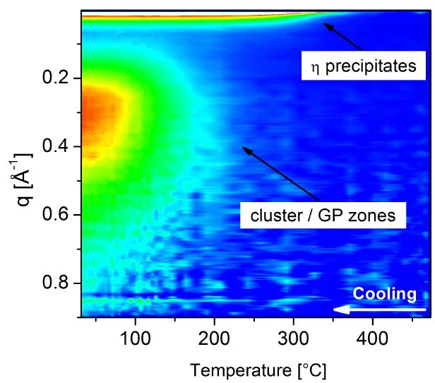
In situ synchrotron x-ray diffraction and small angle scattering are ideal techniques to investigate the kinetics of the microstructure evolution during fast cooling, providing input for models that aim to predict the behaviour of engineering materials under such conditions. More.

In Situ High Resolution Digital Image Correlation

HRDIC

Digital image correlation (DIC) of images obtained during mechanical testing brings possibilities to study localization of plastic deformation on a specimen surface. This technique has been predominantly used in combination with optical cameras or microscopes. Coupling of DIC with a scanning electron microscopy (SEM) enables to obtain significantly more precise data down to the submicron scale. The results of such experiments can provide important data for further design of materials or manufacturing process optimization. More.

Molecular Dynamics Simulations

MD box.jpg

Nanocrystalline (nc) metals are by definition polycrystalline structures with a mean grain size below 100 nm and therefore their microstructure contains a significant volume fraction of interfacial regions separated by nearly-perfect crystals. Compared to its coarse-grain counterpart, the mechanical behavior of a fully-dense nc metal is characterized by a significantly enhanced yield stress and a limited tensile elongation. From the early beginning of experimental investigations in the mechanical behavior of nc metals, it has been proposed that the small grain sizes limit the conventional operation of dislocation sources and estimations of strength were made based on simple Frank–Read sources.

With the increase in computational power, one can deform three-dimensional (3D) computational nc samples containing enough grains in order to mimic an nc metal. These simulations have revealed that the deformation behavior is still to a major extent governed by dislocations: however the dislocations are not nucleated via classical Frank-Read sources situated within the grain interior, but are nucleated at GB ledges, with dislocations traveling through the grain and finally being absorbed in opposite GBs. The merit of the atomistic simulations of nc GB networks lies in the revelation of the details of such dislocation mechanisms. Various examples of our contributions to this research field can be found here.

Crystal Plasticitiy Simulations

Crystal plasticity is a meso-scale modeling approach that predicts the mechanical response of polycrystalline microstructures during plastic deformation. These models may account for the material anisotropy, grain texture and morphology evolution, intergranular and intra-granular stresses and strains, dislocation density kinetics, phase transformations, among others. Crystal plasticity models can be used to gain deeper understanding of the material response that is not accessible via experimental approaches. These models work in synergy with microstructure characterization techniques such as electron backscattering diffraction or in-situ digital image correlation, as well as in-situ diffraction experiments during mechanical testing. We have developed a multi-scale crystal plasticity approach to study multi-axial strain path changes in structural alloys. Details of the model and its application can be found here.

Mit Sidebar

EPFL.png

Contact

PEM
Paul Scherrer Institut
CH-5232 Villigen-PSI
Switzerland

Prof. Helena Van Swygenhoven
Group Leader

Telephone:
+41 56 310 2931
E-mail:
helena.vs@psi.ch

SINQ Facility

Information about the Neutron Source.


LSC Homepage

Laboratory for Condensed Matter Physics


Photon Science Division

Homepage of PSI Division Photon Science (PSD)

Scientific Highlights PSD

Scientific Highlights of PSI Division Photon Science (PSD)

Swiss Light Source SLS

Synchrotron light large research facility.

SwissFEL

The new X-ray free electron laser facility.


User Office

Unterstützung für die externen Nutzer der PSI-Forschungsanlagen


Current openings PSD

Job Opportunities at Research Division Photon Science


Anfahrt zum PSI

Wie Sie uns finden (Beschreibung und Karte)

top

Fussbereich

Paul Scherrer Institut

Forschungsstrasse 111
5232 Villigen PSI
Schweiz

Telefon: +41 56 310 21 11
Telefax: +41 56 310 21 99

Der Weg zu uns
Kontakt

Besucherzentrum psi forum
Schülerlabor iLab
Zentrum für Protonentherapie
PSI Bildungszentrum
PSI Guest House (in english)
PSI Gastronomie
psi forum-Shop


Service & Support

  • Telefonbuch
  • User Office
  • Accelerator Status
  • Publikationen des PSI
  • Lieferanten
  • E-Rechnung
  • Computing
  • Sicherheit

Karriere

  • Arbeiten am PSI
  • Stellenangebote
  • Aus- und Weiterbildung
  • Career Center
  • Berufsbildung
  • PSI Bildungszentrum

Für die Medien

  • Das PSI in Kürze
  • Zahlen und Fakten
  • Mediacorner
  • Medienmitteilungen
  • Social Media

Folgen Sie uns: Twitter (deutsch) LinkedIn Youtube Facebook Instagram Issuu RSS

Footer legal

  • Impressum
  • Nutzungsbedingungen
  • Editoren-Login