
Lab News & Scientific Highlights
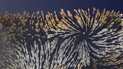
Tauchgang in einen Magneten
Zum ersten Mal haben Forschende die Richtungen der Magnetisierung in einem dreidimensionalen magnetischen Objekt sichtbar gemacht. Die kleinsten Details in ihrer Visualisierung waren dabei zehntausend Mal kleiner als ein Millimeter. In der sichtbar gemachten magnetischen Struktur stach eine Art von Muster besonders hervor: magnetische Singularitäten namens Bloch-Punkte, die bisher nur in der Theorie bekannt waren.
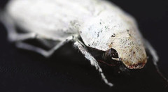
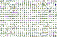
Photonic structure of white beetle wing scales: optimized by evolution
A very thin layer on this beetle’s wings exhibits a complicated structure on the nanoscale that gives them a bright white color. X-ray nanotomography acquired at the Swiss Light Source provides a faithful image of this structure in three dimensions with which scientists can confirm its evolutionary optimization: just enough material for an efficient reflection of white light.
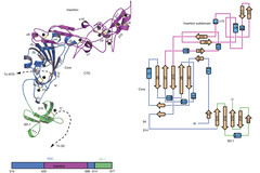
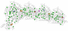
Towards understanding of human betacoronavirus HKU1 life cycle
Researchers from China and USA join forces with Swiss Light Source (SLS) macromolecular crystallography (MX) beamline scientists in a study, which aims at understanding an important step in the life cycle of the human betacoronavirus HKU1.
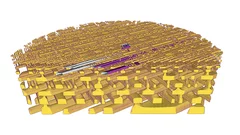
3-D-Röntgenbild macht feinste Details eines Computerchips sichtbar
Forschende des PSI haben detaillierte 3-D-Röntgenbilder eines handelsüblichen Computerchips erstellt. In ihrem Experiment haben sie ein kleines Stück aus dem Chip untersucht, das sie zuvor herausgeschnitten hatten. Diese Probe blieb dabei während der Messung unbeschädigt. Für Hersteller ist es eine grosse Herausforderung, zu bestimmen, ob der Aufbau ihrer Chips am Ende den Vorgaben entspricht. Somit stellen diese Ergebnisse eine wichtige Anwendung eines Röntgen-Tomografieverfahrens dar, das die PSI-Forschenden seit einigen Jahren entwickeln.
1000 Structures solved at X06DA-PXIII
The macromolecular crystallography beamline X06DA-PXIII has reached 1,000 structures in the Protein Data Bank (PDB) on February 22, 2017.
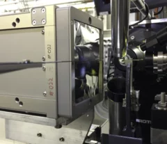
First protein structure solved using the JUNGFRAU detector!
JUNGFRAU is a charge-integrating, two-dimensional pixel detector developed at the Paul Scherrer Institut for use at free-electron lasers, in particular SwissFEL, and synchrotron light sources. On the 10th October, the first protein crystallography experiment using the JUNGFRAU detector, was performed at the beamline X06SA (PXI) of the Swiss Light Source by the members of the Protein Crystallography and Detectors groups at PSI.
Call for expressions of interest: Beamline partners at the SLS for PX II and PX III
We invite companies and institutions to secure access to the beamlines X10SA/PX II and X06DA/PX III through a long term contract.

Single shot grating interferometry demonstrated using direct conversion detection
Researchers at the Paul Scherrer Institute's Swiss Light Source in Villigen, Switzerland, have developed an X-ray grating interferometry setup which does not require an analyzer grating, by directly detecting the fringes generated by the phase grating with a high resolution detector. The 25um pitch GOTTHARD microstrip detector utilizes a direct conversion sensor in which the charge generated from a single absorbed photon is collected by more than one channel. Therefore it is possible to interpolate to achieve a position resolution finer than the strip pitch.
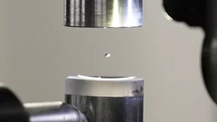
Experiment im schwebenden Tropfen
Der genaue Aufbau von Proteinen wird am PSI standardmässig mittels Röntgenstrahlung entschlüsselt. Nun haben zwei PSI-Wissenschaftler diese Methode trickreich weiterentwickelt: Anstatt die Proteine zu befestigen, untersuchten sie die Proteine in einem frei schwebenden Flüssigkeitstropfen.