
News & Scientific Highlights

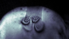
Neurodegenerative disease studied by cryogenic X-ray nanotomography
Hard X-ray cryo-tomography scanning of retina from healthy and inherited blindness specimen paves the way for correlative analysis after imaging at the cSAXS beamline.

Dr. Manuel Guizar-Sicairos is awarded ICO prize
Dr. Manuel Guizar-Sicairos, beamline scientist at the cSAXS beamline, is the 2019 recipient of the International Commission for Optics (ICO) Prize. The distinction was awarded in the EOSAM conference in Rome.
Imaging strain with high resolution
Imaging strain in crystalline materials with high resolution can be a challenging task. Researchers demonstrate an original use of X-ray ptychography for this purpose: ptychographic topography.

Wie Katalysatoren altern
Katalysatoren, die in der Industrie eingesetzt werden, verändern über die Jahre ihre Materialstruktur. Mit einer neuen Methode haben PSI-Forschende dies nun auf der Nano-Skala untersucht.

Quantifying oriented myelin in mouse and human brain
Myelin 'insulates' our neurons enabling fast signal transduction in our brain. Myelin levels, integrity, and neuron orientations are important determinants of brain development and disease. Small-angle X-ray scattering tensor tomography (SAXS-TT) is a promising technique for non-destructive, stain-free imaging of brain samples, enabling quantitative studies of myelination and neuron orientations, i.e. of nano-scale properties imaged over centimeter-sized samples.

Magnetic vortices come full circle
The first experimental observation of three-dimensional magnetic ‘vortex rings’ provides fundamental insight into intricate nanoscale structures inside bulk magnets, and offers fresh perspectives for magnetic devices.

Dr. Manuel Guizar-Sicairos elected as Fellow member of The Optical Society (OSA)
Dr. Manuel Guizar-Sicairos, beamline scientist at the cSAXS beamline, was elected as a Fellow Member of The Optical Society (OSA) for seminal contributions to methods and applications of coherent lensless imaging, ptychography, x-ray nanotomography, and new modalities of x-ray microscopy.

Nanowelten in 3-D
Tomogramme aus dem Inneren von Fossilien, Hirnzellen oder Computerchips liefern neue Erkenntnisse über feinste Strukturen. Die 3-D-Bilder gelingen mithilfe der Röntgenstrahlen der Synchrotron Lichtquelle Schweiz SLS dank eigens entwickelter Detektoren und raffinierter Computeralgorithmen.
Kurzfilm eines magnetischen Nanowirbels
Mit einer neu entwickelten Untersuchungsmethode konnten Forschende die magnetische Struktur im Inneren eines Materials mit Nanometer-Auflösung abbilden. Ihnen gelang ein kurzer «Film» aus sieben Bildern, der erstmalig in 3-D zeigt, wie sich winzige Wirbel der Magnetisierung tief im Inneren eines Materials verändern.