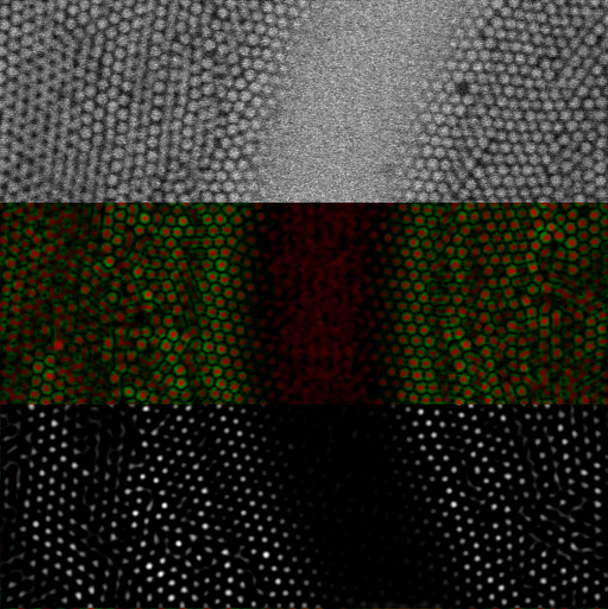
From biology to physics, the range of topics set to benefit from PSI researchers’ improved image analysis for detection of particles is incredibly broad. Publishing their work in the journal Optics Express, the new algorithm improves accuracy and reliability of image analysis by incorporating features found specifically in spherical particles, the most common shape of objects of interest in imaging data.
Quantitative analysis of particles in microscopy images is a topic that ignores disciplinary boundaries. Colloidal suspensions, with particles in the nanometre to micron size scale, big enough to visualise with optical microscopy, are used as models for atomic and molecular systems. Soft matter physicists use them to understand fundamental processes such as how the tiniest crystals from and grow, or, conversely, why some materials remain in a disordered, glassy state. Biologists squint down microscopes to look at biological macromolecules and structures, whether grains of pollen or nanoparticles used as contrast agents or for delivering drugs to cells. In industry, colloids feature in diverse applications, from foods to cosmetics.
However, the fast, accurate and reliable quantitative detection of these particles in images is not straightforward. As images of colloids can contain thousands of particles, automated particle detection algorithms are often used. These particle detection algorithms typically detect a change in brightness, such as at a particle edge. But the images often exhibit a complex background due to diffraction and scattering of light from the spheres. Furthermore, many interesting applications possess heterogeneous sample environments, such as imaging organelles within cellular compartments or studying crystallisation on a surface. Altogether, this results in the generation of numerous false positives or in some of the particles of interest being overlooked.
A more targeted algorithm detects spherical particles
Many colloidal suspensions contain spherical particles. The algorithm, developed by researchers from the Laboratory of Neutron Scattering and Imaging, specifically detects features found in spherical particles.
Whereas traditional methods detect features based on brightness, regardless of shape, their algorithm crucially includes the brightness gradient, thereby adding directional information. “The brightness gradient has a significant magnitude at the edge of the spherical particle and points towards the centre. Using this information in our detection algorithm improves the accuracy and reliability of determined particle positions,” explains researcher, Urs Gasser.
To improve the specificity even further, they then encode the brightness gradient with spherical harmonics, directional functions on the surface of the sphere. Combining these magnifies the directional information to give improved sensitivity in detection of spherical features.
Adding to the toolkit of techniques, the researchers also improved upon another image problem: ‘bridges’. Images of dense colloidal suspensions often show an artefact of bright connections or ‘bridges’ between neighbouring particles, which can reduce accuracy of calculated particle coordinates.
With these changes, the researchers were able to improve the reliability of finding all wanted particles in a microscopy image by one – or even two – orders of magnitude. A large number of unwanted features, detected by other algorithms, could be excluded. Furthermore, the accuracy of calculated particle coordinates were improved.
A key advantage of the new method is speed. Although extremely reliable and accurate methods exist for full image reconstruction, requiring thousands of fit parameters, analysis can take hours. The method devised by PSI researchers provides high quality results in a matter of seconds. For instances, where full image reconstruction is necessary, their algorithm is useful for generating high quality input parameters.
The researchers hope that their algorithm, which enables unwanted features to be excluded, will be helpful to researchers studying particles in heterogeneous sample environments or near to surfaces across the disciplines.
Progress in this field can have big impact, because the applications are so broad. We are interested in the fundamental building blocks in soft matter, but our work is useful for anyone studying spherical particles in microscopy images – no matter what they are or how the image was acquired,
says Urs Gasser.
Text: Miriam Arrell
Contact
Dr. Urs Gasser
Laboratory for Neutron Scattering and Imaging
Paul Scherrer Institut
5232 Villigen
Switzerland
urs.gasser@psi.ch
Original Publication
Urs Gasser and Boyang Zhou
Accurate detection of spherical objects in a complex background
Optics Express 29(23) 37048-37065 (2021)
https://doi.org/10.1364/OE.434652


