Abstract:
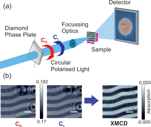
Imaging the magnetic structure of a material is essential to understanding the influence of the physical and chemical microstructure on its magnetic properties. Magnetic imaging techniques, however, have been unable to probe three-dimensional micrometer-size systems with nanoscale resolution. Here we present the imaging of the magnetic domain configuration of a micrometer-thick FeGd multilayer with hard x-ray dichroic ptychography at energies spanning both the Gd L3 edge and the Fe K edge, providing a high spatial resolution spectroscopic analysis of the complex x-ray magnetic circular dichroism. With a spatial resolution reaching 45nm, this advance in hard x-ray magnetic imaging is a first step towards the investigation of buried magnetic structures and extended three-dimensional magnetic systems at the nanoscale.
Facility: SLS; LMX, Mesoscopic Systems
Reference: C. Donnelly et al, Phys. Rev. B 94, 064421 (2016)
Read full article: here

