Aller au contenu principal
  • Paul Scherrer Institut PSI
  • PSI Research, Labs & User Services

Digital User Office

  • Digital User Office
  • DE
  • EN
  • FR
Paul Scherrer Institut (PSI)
Rechercher
Paul Scherrer Institut (PSI)

Hauptnavigation

  • Research at PSIOuvrir ce point de menu principal
    • Research Initiatives
    • Research Integrity
    • Scientific Highlights
    • Scientific Events
    • Scientific Career
    • PSI-FELLOW
    • PSI Data Policy
  • Research Divisions and LabsOuvrir ce point de menu principal
    • Overview
    • Research with Neutrons and Muons
    • Photon Science
    • Energy and Environment
    • Nuclear Energy and Safety
    • Biology and Chemistry
    • Large Research Facilities
  • Facilities and InstrumentsOuvrir ce point de menu principal
    • Overview
    • Large Research Facilities
    • Facilities
    • PSI Facility Newsletter
  • PSI User ServicesOuvrir ce point de menu principal
    • User Office
    • Methods at the PSI User Facilities
    • Proposals for beam time
    • Proposal Deadlines
    • Data Analysis Service (PSD)
    • EU support programmes
  • DE
  • EN
  • FR

Digital User Office (mobile)

  • Digital User Office

Vous êtes ici:

  1. PSI Home
  2. Labs & User Services
  3. PSD
  4. LMN
  5. Research Groups
  6. SwissFEL nanoengineering
  7. Supports for Serial Protein Crystallography

Navigation secondaire

Laboratory for Micro and Nanotechnology

  • About LMN
    • Organisational Structure
  • Open Positions
  • People
  • Research Groups Sous-menu élargi
    • Nanotechnology
    • X-ray Optics and Applications
      • X-ray Optics for Imaging and Spectroscopy
        • Fresnel Zone Plate for X-ray Microscopy
        • Blazed X-ray Optics
        • Zernike X-ray Phase Contrast Microscopy
        • Fresnel Zone Plates for RIXS
        • Refractive Lenses by 2 Photon 3D Lithography
      • Wavefront Metrology and Manipulation
        • Vortex Fresnel Zone Plates
        • Grating-based Wavefront Metrology
      • X-ray Optics for XFELs
        • Diamond Fresnel Zone Plates
        • Beam Splitter Gratings for Spectral Monitoring
        • A Delay Line for Ultrafast Pump-Probe Experiments
        • X-ray Streaking for Ultrafast Processes
    • Polymer Nanotechnology
      • Nanoimprint Lithography
      • Three Dimensional Structures
    • Molecular Nanoscience
      • On-surface Chemistry
      • Spins in Molecular Monolayers
      • SiC: Surfaces and Interfaces
      • Our Research Team
    • Advanced Lithography and Metrology
      • EUV Interference Lithography
      • EUV Lensless Imaging
      • ALM Nanoscience
    • Quantum Technologies
      • News and highlights
      • People
      • Open positions
      • Current projects
        • 2D semiconductor devices
        • CDW-based memory devices
        • Imaging quantum many-body states
        • Nonlinear magnonics
        • Rare-earth quantum magnets
        • Strained Germanium laser
      • Techniques
        • Cristallina-Q
        • IR beamline
        • Nano-fabrication
      • Publications
      • QTC@PSI
  • Facilities and Equipment
    • Cleanroom Labs
    • Surface Science Lab
    • Scanning Electron Microscopy
    • Scanning Probe Microscopy
    • PEARL Beamline
    • XIL Facility at the SLS
    • Nanoimprint Facilities
    • Electron Beam Lithography
  • LMN News
  • LMN Highlights
    • Archive
  • Publications
    • Publications 2011 - 2016

Info message

Ce contenu n'est pas disponible en français.

Solid supports for X-FEL based dynamic studies on 2D and 3D nanocrystals of membrane proteins.

In a joint project with the Center for Cellular Imaging and NanoAnalytics (C-CINA) at the University of Basel and the Lab. for biomolecular research at PSI we are developing tools and methods for the preparation of protein crystals for dynamic studies at XFEL sources. The project is financed by the Swiss Nanoscience Institute.


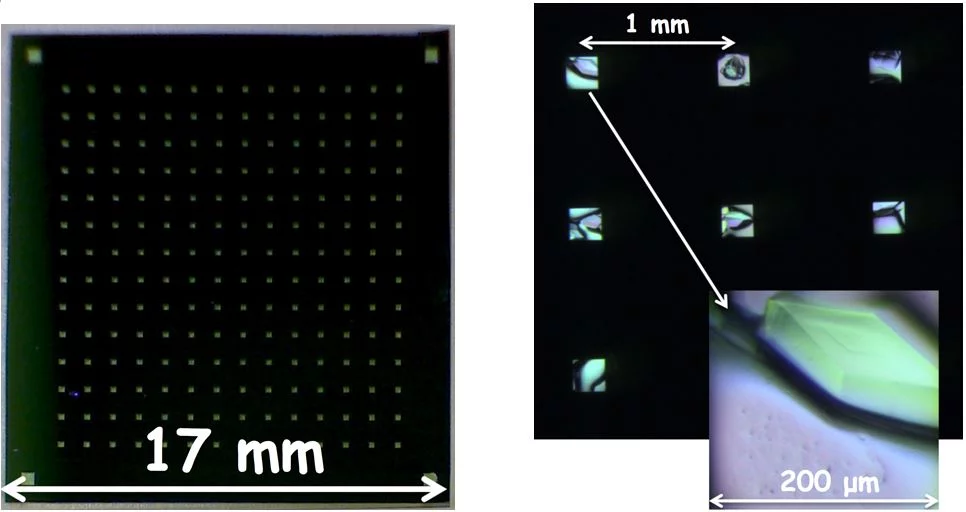
Lysozyme crystals grown in situ on silicon nitride window supports.
Serial femtosecond crystallography based on X-ray free-electron laser sources (XFELs) provides new opportunities for structural sciences, in particular for the time-resolved investigation of dynamic processes [1-3]. Even though after every ultrafast shot of a very intense coherent laser pulse the sample is destroyed due to Coulomb explosion, X-ray diffraction data can be collected in the so-called ‘diffract-before-destroy’ regime.

High efficiency measurements at XFELs require high density and very well ordered deposition of the biological sample – usually two dimensional (2D) or three dimensional (3D) crystals of proteins – on a substrate. The mount, which has an array of ultrathin windows (Fig. 1), is positioned on a moveable stage allowing it to be scanned by the (fixed) laser beam. This approach gives much higher hit rates and much lower sample consumption than the liquid jets commonly used to inject a large number of small crystals into the beam.

Microfabricated solid supports (fixed targets) filled with crystals protected from dehydration, allow measurements to be performed at room temperature, enabling structure determination of macromolecules in a close-to-native environment at high resolution [4].

Well-established microfabrication technology based on materials such as silicon and silicon nitride makes it possible to create ultrathin, low background packaging, that is especially useful for supporting 3D nanocrystals or 2D crystals, which have too low diffraction yields for reasonable measurements to be made using synchrotron radiation.

Further development towards highly X-ray transparent, less fragile, easy to mass-fabricate devices is required to allow future cost-efficient high throughput measurements at FELs. The main aim of this project is to improve sample preparation methods for XFEL-based protein nanocrystallography. In particular, we develop supports optimized for protein crystal preparation, to minimize sample consumption by adapting the liquid handling system and to perform proof-of-concept pump-probe experiments on solid supports [5,6].

References:
[1] I. Schlichting, IUCrJ, 2015, 2, 246-255
[2] M. Frank et al., IUCrJ, 2014, 1, 95–100
[3] S. Boutet et. al, Science 337, 2012, 362
[4] A. Zarrine-Afsar et al., Acta Cryst. 2012, D68, 321-323
[5] N. Opara et al., "Direct protein crystallization on ultrathin membranes for diffraction measurements at X-ray free electron lasers." J. Appl. Cryst. (2017) 50, 909-918.
[6] N. Opara et al., "Demonstration of femtosecond X-ray pump X-ray probe diffraction on protein crystals". Structural Dynamics 5, 054303 (2018).


Sidebar

Contact

Dr. Celestino Padeste

Division of Biology and Chemistry
Paul Scherrer Institut
Forschungsstrasse 111
5232 Villigen PSI
Switzerland

Telephone:
+41 56 310 2141

E-mail:
celestino.padeste@psi.ch

top

Pied de page

Paul Scherrer Institut

Forschungsstrasse 111
5232 Villigen PSI
Suisse

Téléphone: +41 56 310 21 11
Téléfax: +41 56 310 21 99

Comment nous trouver 
Formulaire de contact

Centre de visiteurs psi forum
Laboratoire élèves iLab (en allemand)
Centre de protonthérapie
Centre de Formation du PSI (en allemand)
PSI Guest House (en anglais)
PSI Gastronomie (en allemand)

Service & Support

  • ​Annuaire/Liste de contacts
  • User Office
  • Accelerator Status
  • Publications du PSI
  • Fournisseurs
  • E-facture
  • Computing (en anglais)
  • Sicherheit (en allemand)

Carrière

  • Travailler au PSI
  • Offres d'emploi
  • Formation initiale et formation continue
  • Formation professionnelle (en allemand)
  • Centre de Formation du PSI

Pour les médias

  • Le PSI en bref
  • Chiffres et faits
  • Contact destiné aux médias
  • Communiqués de presse
  • Social Media Newsroom

Suivez le PSI: Twitter (en allemand) LinkedIn Youtube Issuu RSS

Footer legal

  • Impressum
  • Conditions d'utilisation
  • Login éditeurs