Hearing loss is a relevant condition worldwide, severely affecting patients of all ages in their quality of life. The WHO identifies 466 million people with disabling hearing loss, 34 million are children, and the tendency is rising [1]. Many cases are conductive hearing loss, which occurs when the sound passage is blocked in either the ear canal or the middle ear.
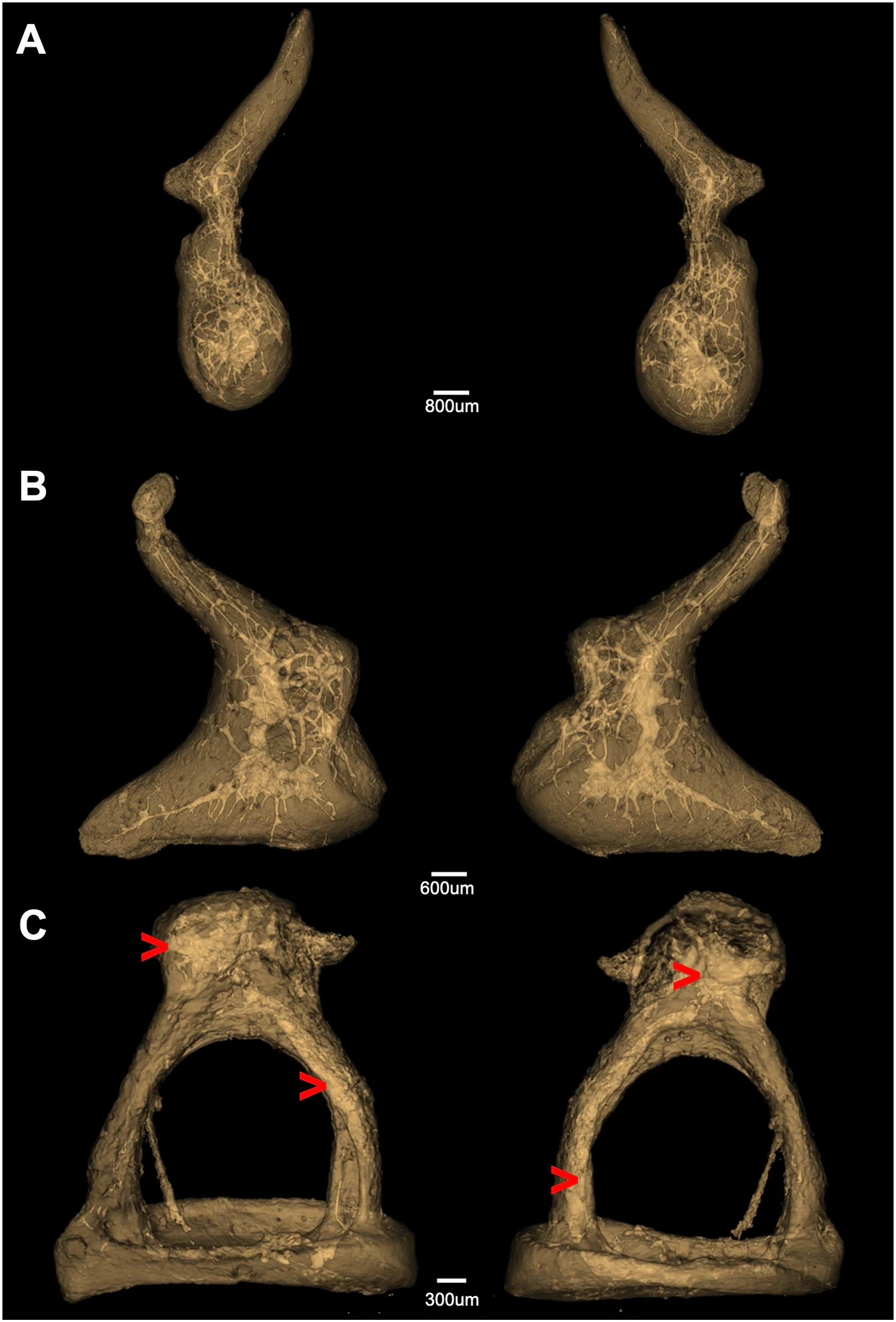
The transmission of sound from the environment to the inner ear's sensory cells depends on the external auditory canal, the tympanic membrane, and the middle ear. The middle ear is an air-filled space that contains the ossicles (malleus, incus, and stapes), which transmit and amplify incoming sound waves from the air to the inner ear. Although the middle ear has been subject to extensive research, middle ear biomechanics are poorly understood and challenging to study. This is mainly due to the involved anatomical structures' size and location. Currently established methods to investigate middle ear biomechanics, like Laser Doppler Vibrometry (LDV), are subject to considerable limitations, [2-4]. Additionally, the motions of interest are tiny, e.g., for a 140 dB SPL stimulus, the displacement of the stapes footplate is, on average, 10 um [2]. To overcome the existing limitations, the TOMCAT beamline offers advanced and unique capabilities to meet the temporal and spatial resolution requirements for middle ear imaging, which have successfully been shown to visualize the sub-micron structure of the human auditory ossicles [5] (see figure). In this project, we will apply synchrotron-based dynamic phase-contrast microtomography as an imaging method that enables the study of the human ossicles' micromotions in a non-destructive manner in their natural, three-dimensional full configuration. The proposed technique has the potential to revolutionize the understanding of middle ear biomechanics. With a detailed understanding of the ossicles' micromotions we could provide significant findings and surgical implications to optimize passive or active middle ear implants (e.g., piston implant as stapes prosthesis).
The project entitled “The Human Auditory System in Motion: Direct Observation of the Microfunction of Sound Transmission using Dynamic Phase-contrast X-ray Imaging” will be done in collaboration with the Inselspital and the ARTORG Center for Biomedical Engineering Research in Bern and is financed by the Swiss National Fund (SNF). Dr. Anne Bonnin is co-PI and the official project leader at PSI.
References
- https://www.who.int/news-room/fact-sheets/detail/deafness-and-hearing-loss (accessed 16.09.2020)
- Hato N, Stenfelt S, Goode RL. Three-dimensional stapes footplate motion in human temporal bones. Audiol Neurootol. 2003 May-Jun;8(3):140-52. PMID: 12679625.
- Lauxmann, Michael & Eiber, Albrecht & Heckeler, Christoph & Ihrle, Sebastian & Chatzimichalis, M & Huber, Alex & Sim, J. (2012). In-plane motions of the stapes in human ears. The Journal of the Acoustical Society of America. 132. 3280-91.
- Anschuetz, L., Demattè, M., Pica, A., Wimmer, W., Caversaccio, M., & Bonnin, A. (2019). Synchrotron radiation imaging revealing the sub-micron structure of the auditory ossicles. Hearing Research, 383, 107806 (8 pp.).
Collaboration
- PD Dr. med. Lukas Anschütz, ENT Department, Inselspital Bern
- Prof. Dr. med. Marco Caversaccio, Director and Chairman ENT Department, Inselspital Bern
- Dr. Wilhelm Wimmer, Group leader Hearing Research Laboratory (HRL), ARTORG Center, University of Bern
Funding
Swiss National Foundation (SNF) - Nr. 320030 192660
Contact
- Dr. Aleksandra Ivanovic, aleksandra.ivanovic@psi.ch
- Dr. Margaux Schmeltz, margaux.schmeltz@psi.ch
