

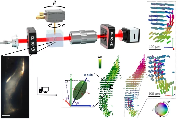
The 3D characterization of ultrastructure, nanoscales structures too small to be imaged using standard light microscopy approach, is key to understand hierarchical materials and their functional properties. We developed a new method, tomographic Müller-polarimetric microscopy (TMPM), that allows to retrieve at three-dimensional microscopic resolution the nanoscale structural information of the ultrastructure probed with polarized light in a non-destructive manner using a low cost and experimentally simple optical setup.
© Paul Scherrer Institute / Arthur Baroni & Yang Chen
The method retrieves the 3D polarimetric properties of thick sample, here modelled as a cluster of uniaxial crystal elements, by combining Mueller polarimetry with the concept of tensor tomography. In this study we demonstrate the method on a on human trabecular bone retrieving its structural information such as orientation of collagen fibers and their degree of orientation, and we conclusively compared the reconstructed 3D arrangement with small-angle X-rays tensor tomography.
Being able to perform these measurements with visible light in a non-destructive manner will enable the study of small model organisms in vivo following development stages or progress of diseases or allows for undisturbed in- situ mechanical tests.
We believe that the technique has a wide range of applications, in particular in biology, medicine and life science. The experimental setup is comparably cheap and easy to implement, and we provide an open-access software package MUMOTT (mumott.org) for the computational reconstruction.
© Paul Scherrer Institute / Atreyee Acharya
Contact
Dr. Arthur Baroni
Structure and Mechanics of Advanced Materials, Center for Photon Science
Paul Scherrer Institute, Forschungsstrasse 111, 5232 Villigen PSI, Switzerland
Telephone: +41 56 40 44, e-mail: arthur.baroni@psi.ch
Dr. Marianne Liebi
Structure and Mechanics of Advanced Materials, Center for Photon Science
Paul Scherrer Institute, Forschungsstrasse 111, 5232 Villigen PSI, Switzerland
Telephone: +41 56 310 44 38, e-mail: marianne.liebi@psi.ch
Original Publication
Reconstructing three-dimensional optical anisotropy with tomographic Müller-polarimetric microscopy. Yang Chen*, Arthur Baroni*, Torne Tänzer, Leonard Nielsen, Marianne Liebi*, Adv. Sci. 2025, 2502075.
*contact authors
Acknowledgements
This work was financed by the European Research Council Starting Grant MUMOTT (ERC-2020-StG 949301), funded by the European Union. Views and opinions expressed are however those of the author(s) only and do not necessarily reflect those of the European Union or the European Research Council Executive Agency. Neither the European Union nor the granting authority can be held responsible for them.