

This home-built free space setup aims at measuring the polarizance of the sample in 3D. To do so, it performs multiple Mueller polarimetric acquisition (see setup Mueller Polarimetric Microscope) at different view of the sample in a tomographic fashion, i.e. at different tilts and rotation of the sample. Due to the use of visible light, the sample need to be thin enough to be always in focus and is therefore often milled into a small cylinder shape (~200 um). To remove unwanted edge scattering and lens effect, the sample is immersed in matching index liquid. Using a gradient based iterative algorithm, one can retrieve from those measurements the 3D birefringence properties of the sample, in its full volume. See https://doi.org/10.1002/advs.202502075
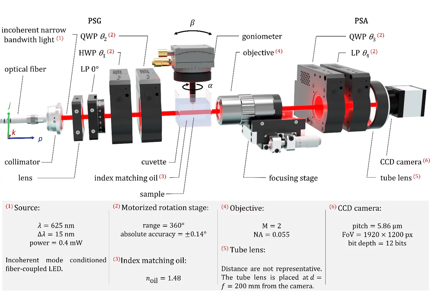
© Paul Scherrer Institute / Arthur Baroni
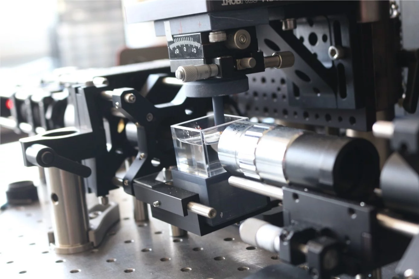
© Paul Scherrer Institute / Arthur Baroni
Example of results
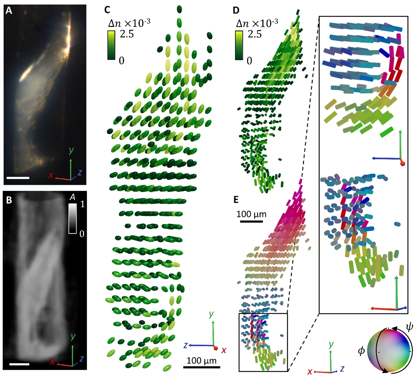
© Paul Scherrer Institute / Arthur Baroni & Yang Chen