In the context of the SLS2.0 upgrade project, the possibility to gain information on the elemental distribution, in addition to the microstructure, in a rapid manner is explored. We aim at combining energy resolved 2D detectors (e.g. Mönch) with pinholes in a camera obscura geometry. We are currently assessing the achievable spatial, temporal and energy resolution as well as the sensitivity of such a system, and developing the required reconstruction and data processing tools. Finally, the most suitable applications are explored.
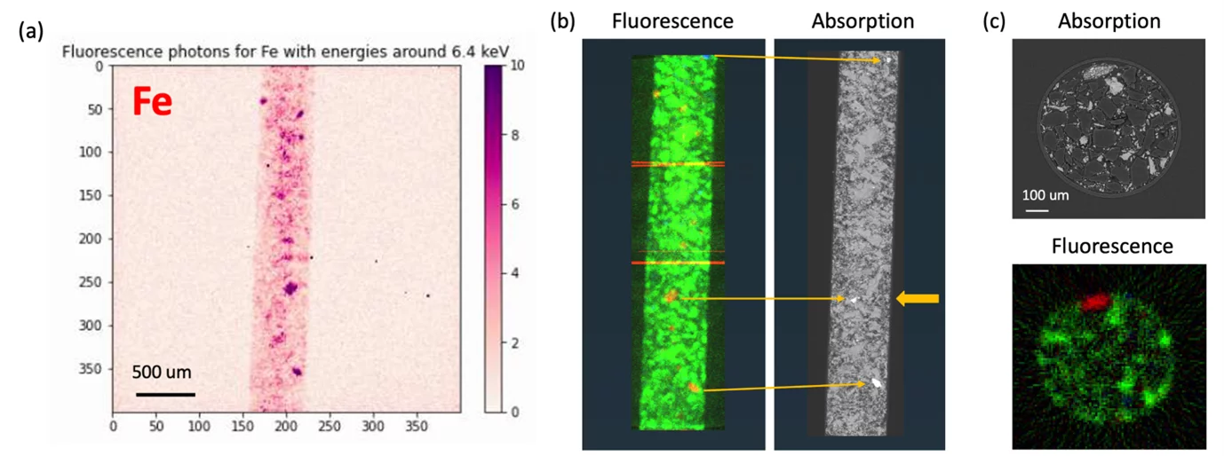
Preliminary results of a proof-of-principle full-field XRF tomographic experiment performed at TOMCAT. The sample was a glass capillary filled with boron nitride beads and grains of malachite (Cu – green in (b-c)), pyrite (Fe – red in (b-c)), hematite (Fe – red in (b-c)) and yttrium oxide (Y – blue in (b)) - (a) 2D projection of the fluorescence photons with an energy of ca. 6.4 keV, (b) Maximum intensity projections of the reconstructed 3D volumes based on fluorescence and absorption, (c) Tomographic slices through the 3D volumes at the position indicated by the fat orange arrow in (b).
Collaboration
- Dr. Anna Bergamaschi, Detector Group, Paul Scherrer Institut
- Dr. Dario Ferreira Sanchez, microXAS Beamline, Paul Scherrer Institut
Contact