
Scientific Highlights and News
The upgrade of the SLS to a diffraction-limited storage ring (DLSR) will benefit in particular those experimental techniques that exploit coherence and/or beam collimation and tight focussing. The increased coherent fraction, of the order of several percent in the hard x-ray regime, will greatly enhance phase-contrast tomography and lensless-imaging techniques such as ptychography; the ability to focus down to micron dimensions while maintaining excellent collimation will allow the investigation of proteins that only form micro- and nanocrystals, most notably membrane proteins and G-coupled protein receptors (GCPRs). The below articles contain recent news and examples, all performed at the SLS, which have been selected as representatives of some of the clearest scientific drivers for the upgrade of the SLS; such experiments at SLS 2.0 will be able to be performed either much more rapidly, or with significantly greater spatial resolution.
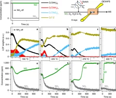
Time-resolved copper speciation during selective catalytic reduction of NO on Cu-SSZ-13
Through the combination of time-resolved X-ray absorption spectroscopy and transient experimentation, we were able to capture an ammonia inhibition effect on the rate-limiting copper re-oxidation at low temperature.
Nanostructure surveys of macroscopic specimens by small-angle scattering tensor tomography
The mechanical properties of many materials are based on the macroscopic arrangement and orientation of their nanostructure. This nanostructure can be ordered over a range of length scales. In biology, the principle of hierarchical ordering is often used to maximize functionality, such as strength and robustness of the material, while minimizing weight and energy cost.
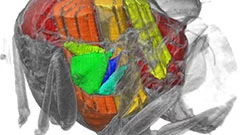
Un film en 3D montre ce qui se passe à l’intérieur d’insectes en plein vol
Grâce aux rayons X produits par la Source de Lumière Suisse SLS, des prises de vue inédites des muscles que les mouches utilisent pour voler (muscles alaires) ont pu être réalisées, à haute vitesse et en 3D. Une équipe de scientifiques de l’Université d’Oxford, de l’Imperial College de Londres et de l’Institut Paul Scherrer (PSI) a développé un procédé de prise de vue tomographique révolutionnaire. Grâce à lui, ils ont pu filmer ce qui se passe à l’intérieur d’insectes en plein vol. Ces films permettent de découvrir en profondeur l’un des mécanismes naturels les plus complexes, et montrent que les déformations structurelles sont la clé pour comprendre la manière dont la mouche contrôle son battement d’aile.