
Show filters
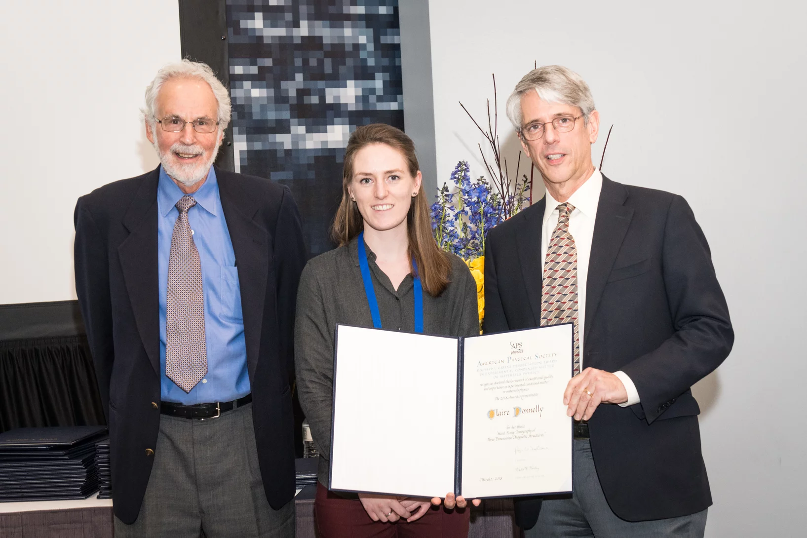
Claire Donnelly dissertation research awards
Claire Donnelly, Mesoscopic Systems (ETH Zurich - PSI), was awarded the COMSOL SPS Award in Computational Physics, the Werner Meyer-Ilse Memorial Award, the ETH Medal for an outstanding doctoral thesis, and the American Physical Society Richard L. Greene Dissertation Award.
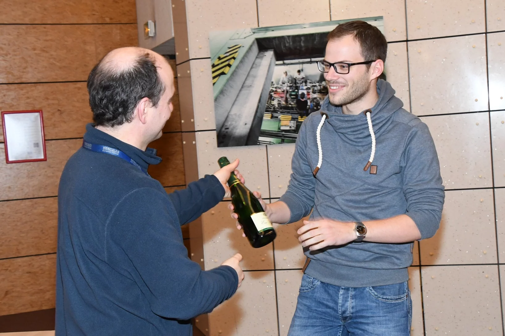
HERCULES School Poster Prize
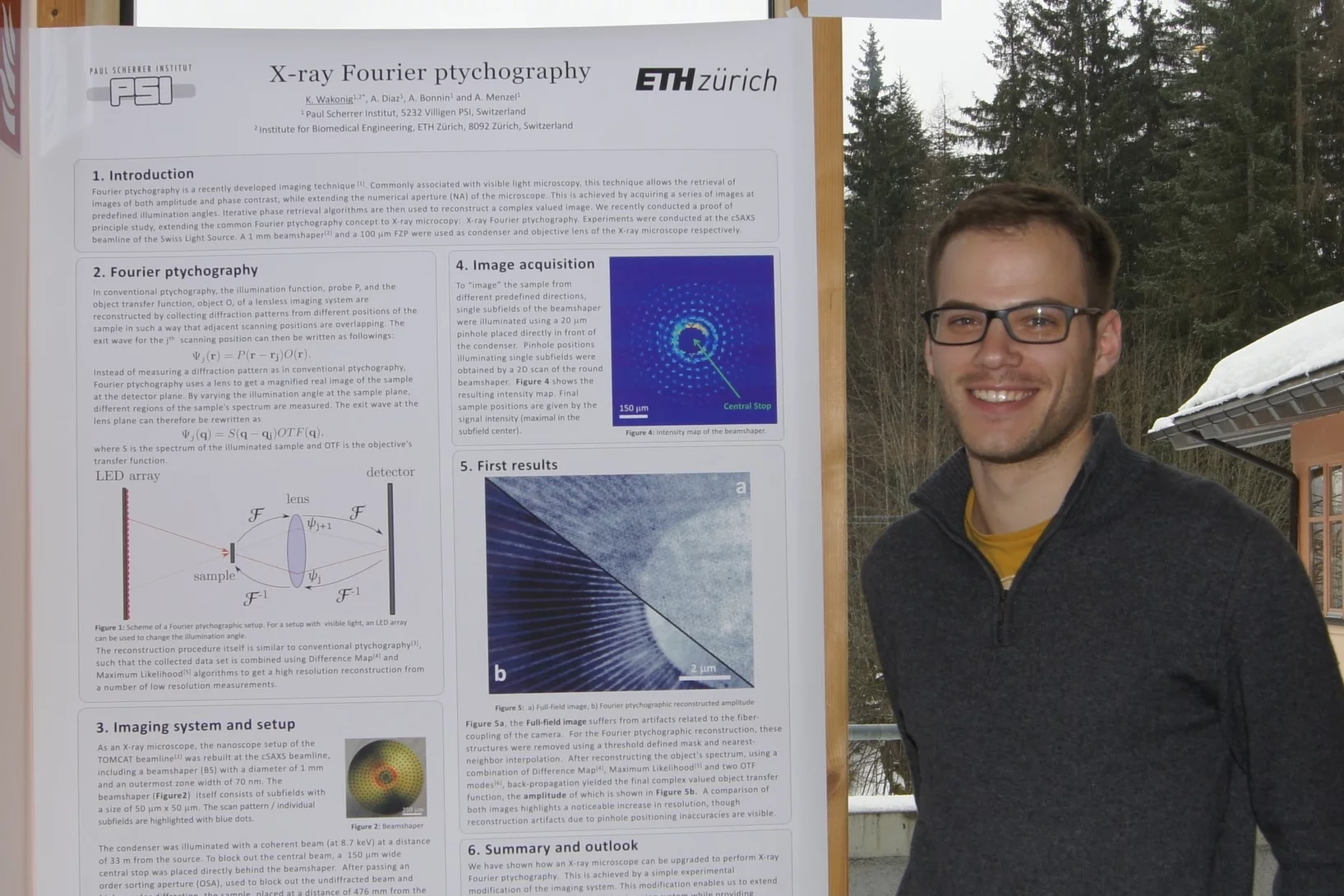
Klaus Wakonig was awarded the best poster prize in the 2018 rendition of the HERCULES European School in Grenoble, France. Klaus is currently a PhD student at the CXS group at PSI, developing X-ray Fourier ptychography.
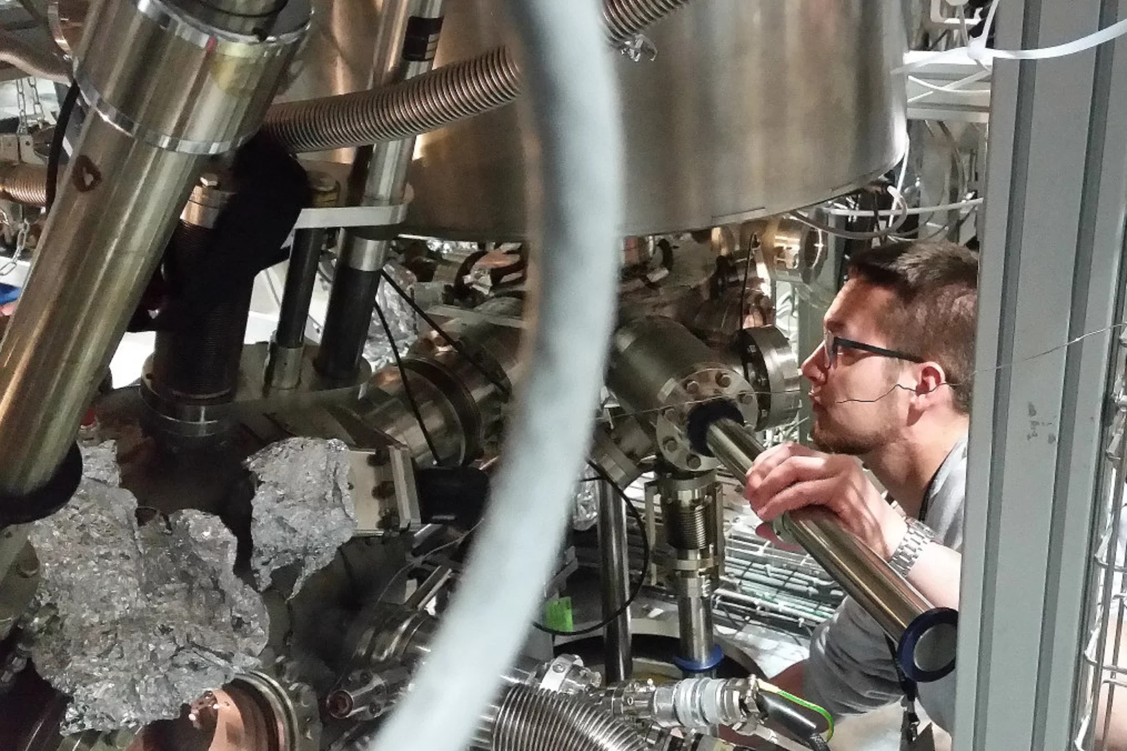
HERCULES at the Swiss Light Source
In the week of March 18-23 PSI welcomes 20 PhD students and postdocs taking part in the HERCULES 2018 school on Neutron and Synchrotron Radiation. They will attend lectures and perform two days of practical courses at several beam lines of the Swiss Light Source.
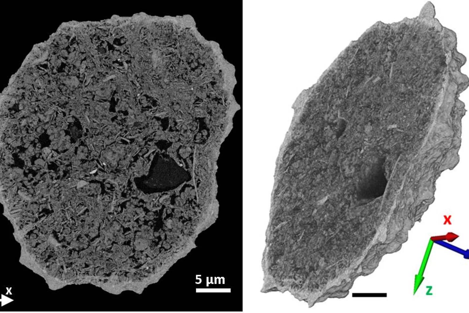
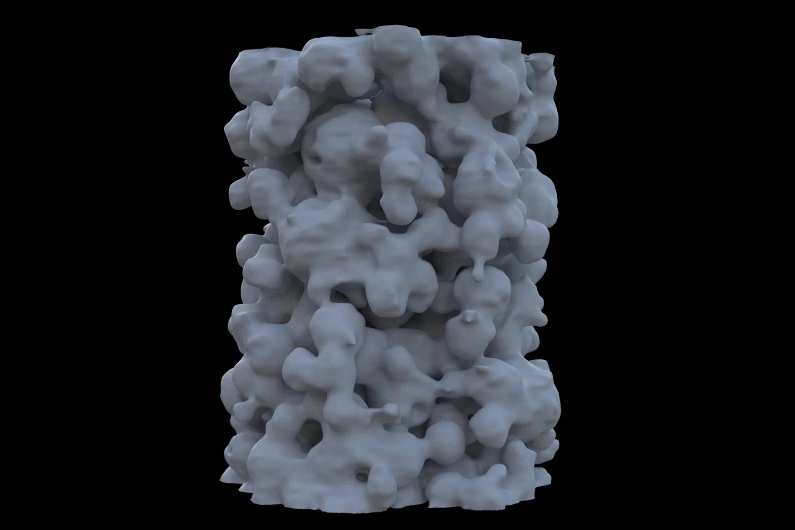
Making the world go round - a look into the structure of a prominent heterogeneous catalyst
Fluid catalytic cracking catalysts, which are composite particles of hierarchical porosity, were examined using ptychographic X-ray tomography. These particles are essential to the conversion of crude oil into gasoline. Examination of catalysts at decreasing levels of catalytic conversion efficacy allowed the detection of possible deactivation causes.
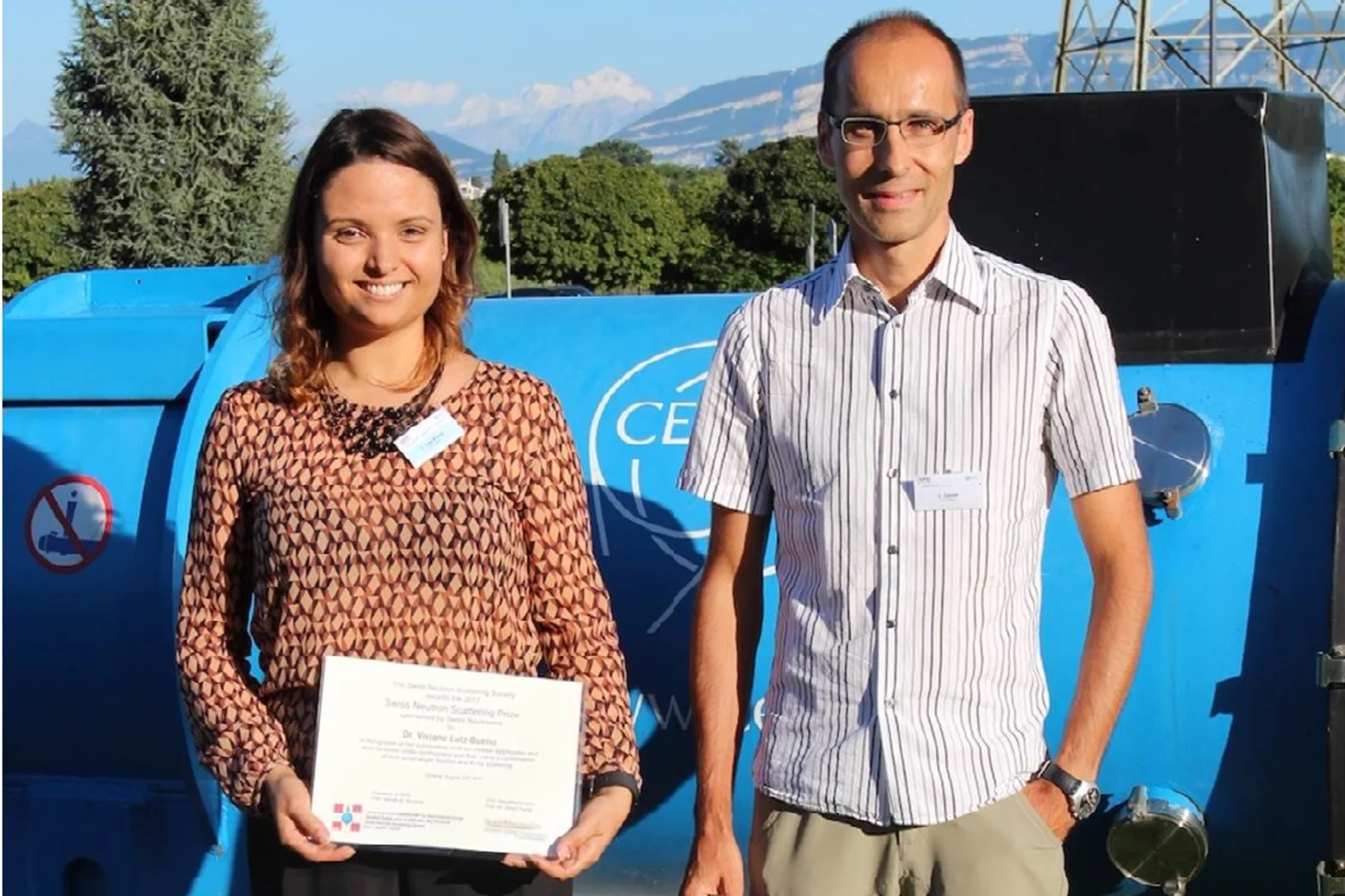
Swiss Neutron Scattering Prize
Viviane Lutz-Bueno was awarded the Swiss Neutron Scattering Prize of the Swiss Neutron Scattering Society, at the Joint Annual Meeting of the Swiss and Austrian Physical Society, for her PhD thesis Effects of formulation and flow on the structure of micellar aggregates carried out at ETH Zürich. Viviane is currently a postdoctoral fellow at the CXS group at PSI, developing scanning SAXS analysis methods.
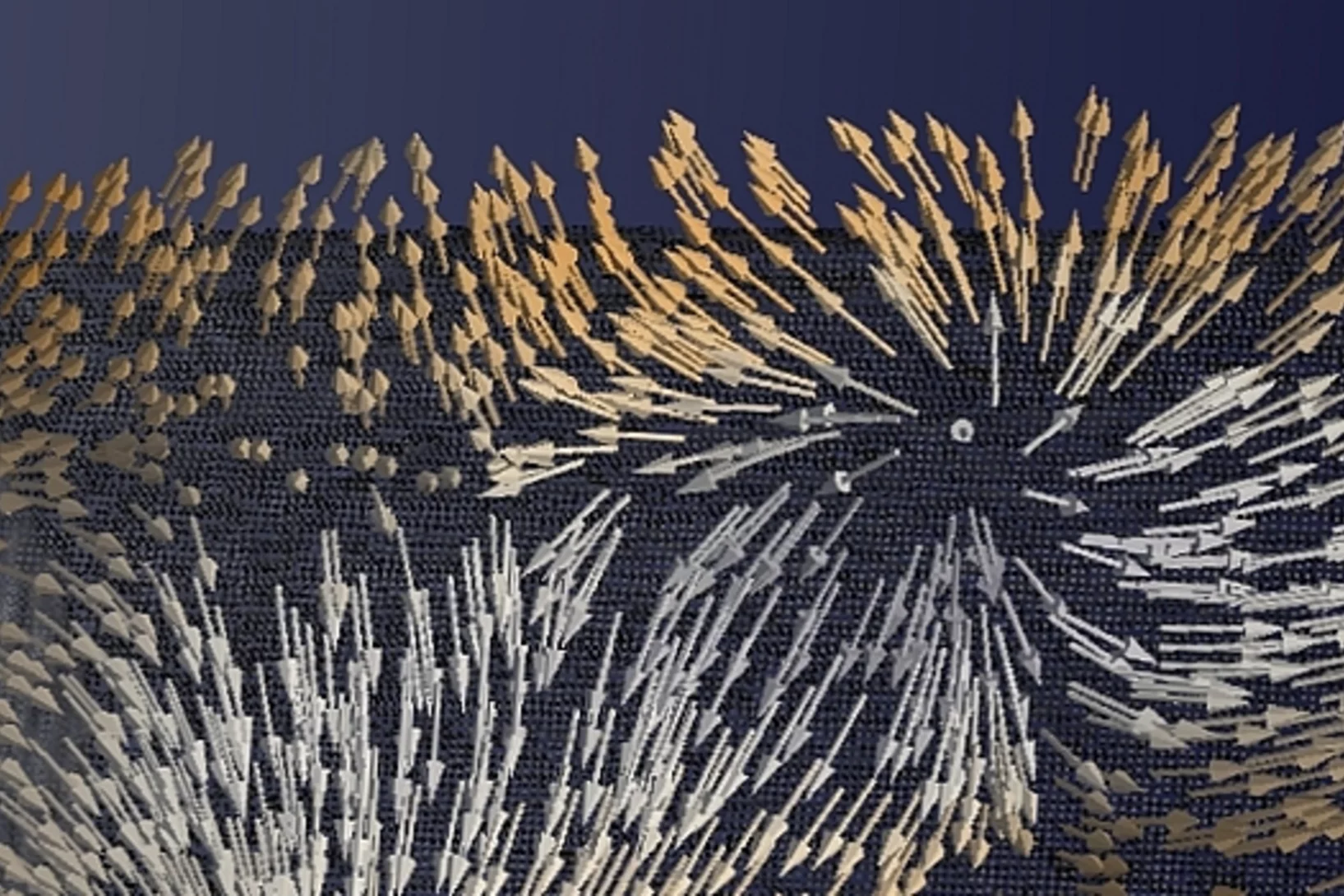
Plongée dans un aimant
Pour la première fois, des chercheurs ont réussi à visualiser les directions de l'aimantation dans un objet magnétique tridimensionnel. Les plus petits détails de leur visualisation mesuraient moins d'un dixième de millième de millimètre. Un type de motif exceptionnel est ressorti dans la structure qu'ils ont fait apparaître: des singularités magnétiques appelées points de Bloch, jusque-là connues uniquement en théorie.
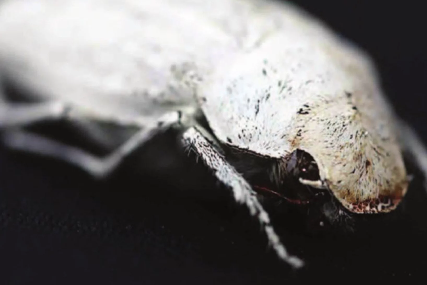
Photonic structure of white beetle wing scales: optimized by evolution
A very thin layer on this beetle’s wings exhibits a complicated structure on the nanoscale that gives them a bright white color. X-ray nanotomography acquired at the Swiss Light Source provides a faithful image of this structure in three dimensions with which scientists can confirm its evolutionary optimization: just enough material for an efficient reflection of white light.
European NESY Winterschool Young Scientist Best Poster Prize
Klaus Wakonig was awarded the "Young Scientist Best Poster Prize" along with a cash prize at the 10th European NESY Winterschool & Symposium on Neutron and Synchrotron Radiation. Klaus is a PhD student at the coherent X-ray scattering group (CXS) in PSI. His poster, entitled "X-ray Fourier ptychography," details his latest results in the implementation of Fourier ptychography at X-ray wavelengths for nanoimaging. Image credit ©NESY/Montanuniversitaet Leoben
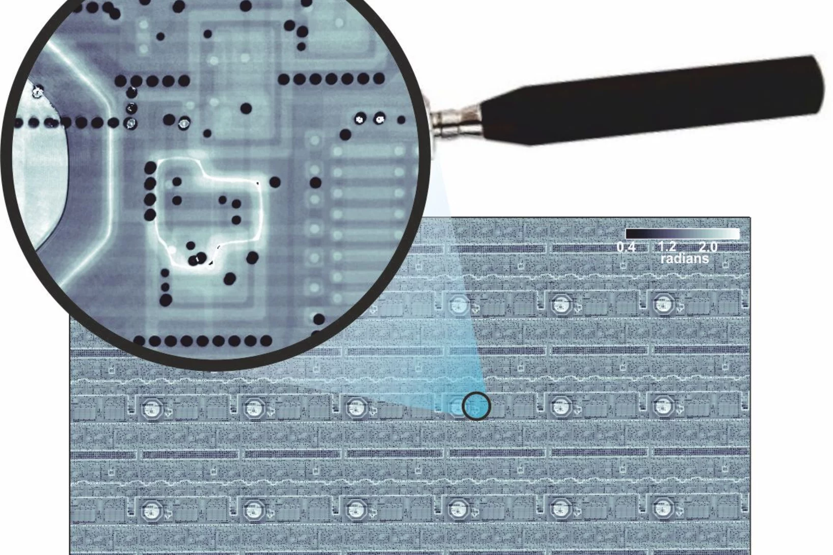
La radiographie en 3D permet de visualiser les moindres détails d’une puce informatique
Des chercheurs de l’Institut Paul Scherrer PSI ont réalisé des radiographies détaillées en 3D d’une puce informatique usuelle. Dans le cadre de leur expérience, ils ont analysé une petite portion de puce qu’ils avaient préalablement découpée. Durant la mesure, cet échantillon est resté intact. Pour les fabricants, déterminer si la structure de leurs puces est conforme aux normes représente un défi. Ces résultats constituent donc une possibilité d’application importante pour un procédé spécifique de tomographie à rayons X que les chercheurs du PSI développent depuis quelques années.
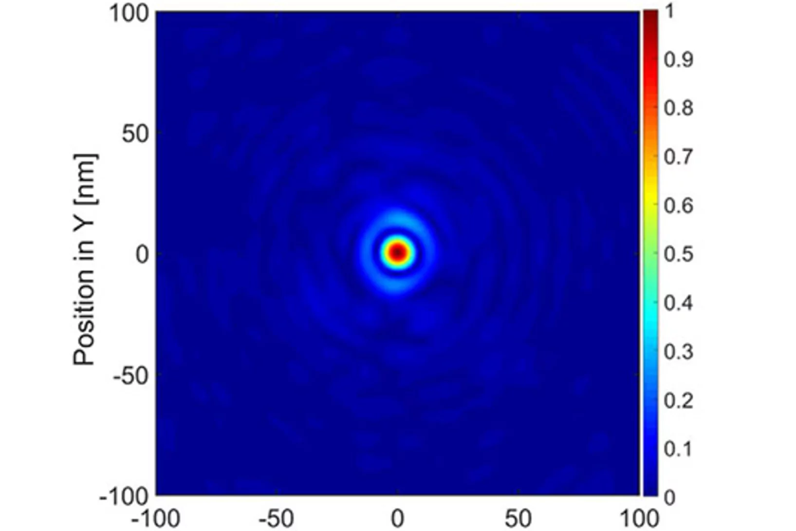
Interlaced zone plates push the resolution limit in x-ray microscopy
A novel type of diffractive lenses based on interlaced structures enable x-ray imaging at resolutions below 10 nm. The fabrication method and the test results of these novel x-ray lenses have been published in the journal Scientific Reports.
How does food look like on the nanoscale?
The answer to this question could save food industry a lot of money and reduce food waste caused by faulty production. Researchers from the University of Copenhagen and the Paul Scherrer Institut have obtained a 3D image of food on the nanoscale using ptychographic X-ray computed tomography. This work paves the way towards a more detailed knowledge of the structure of complex food systems.
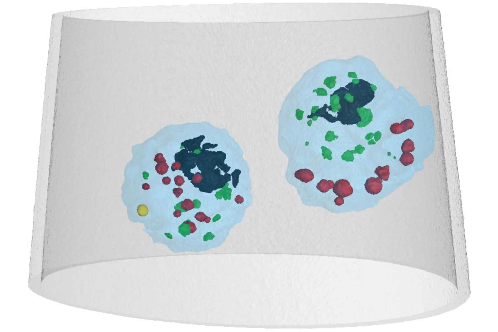
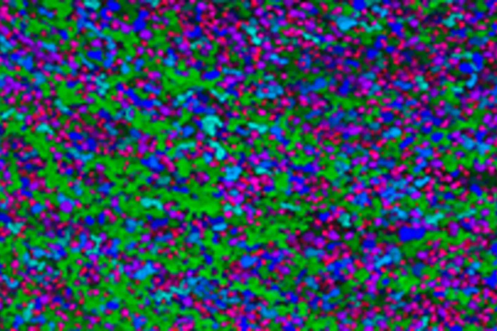
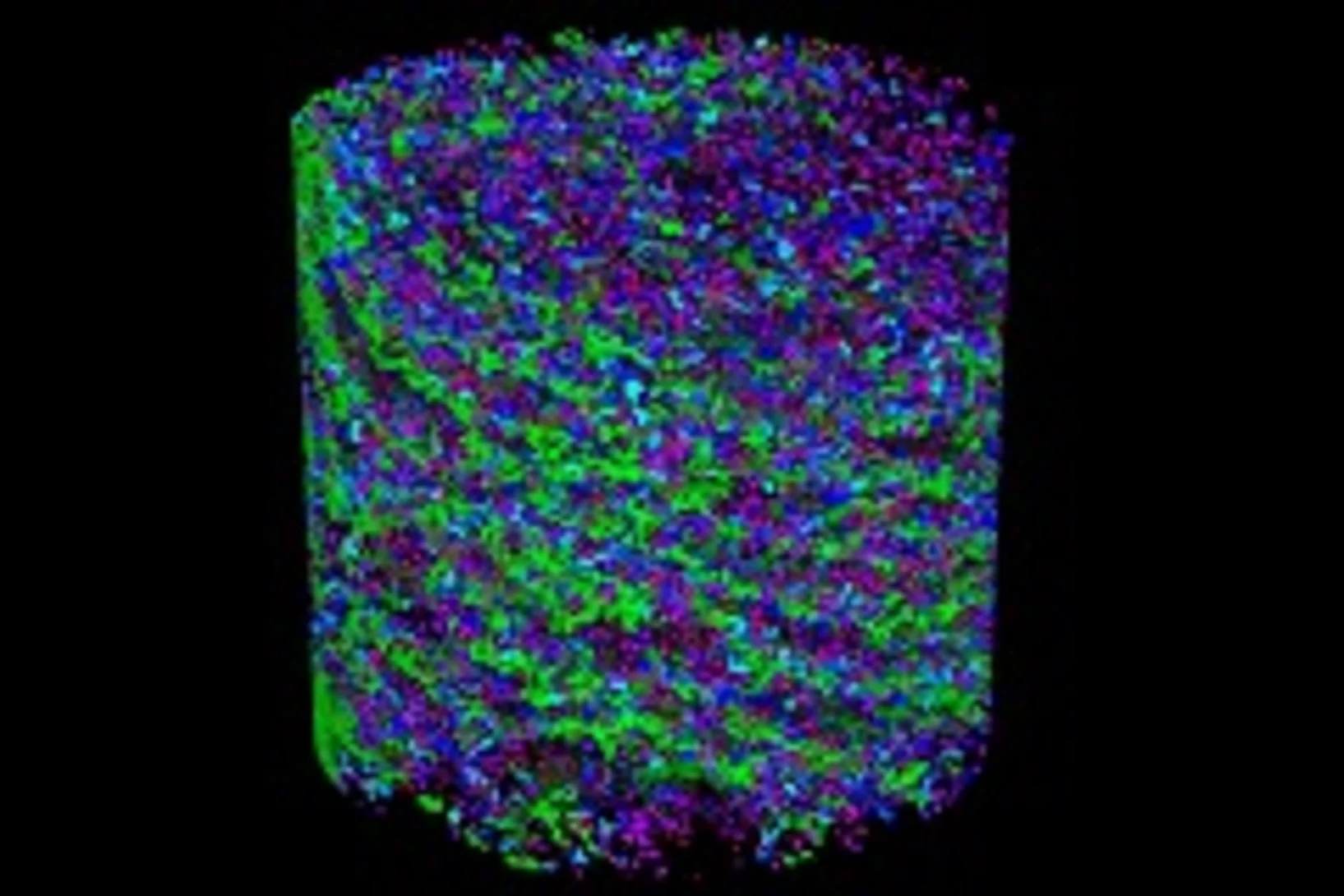
Mass density distribution of intact cell ultrastructure
The determination of the mass density of cellular compartments is one of the many analytical tools that biologists need to unravel the extremely complex structure of biological systems. Cryo X-ray nanotomography reveals absolute mass density maps of frozen hydrated cells in three dimensions.
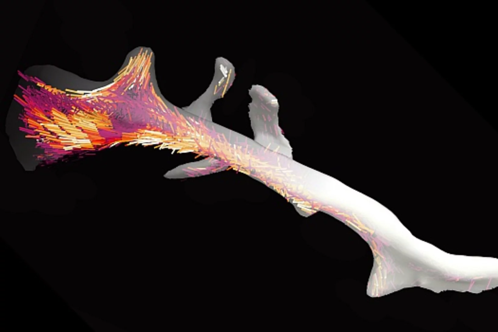
La nanostructure d’un os dévoilée en 3D
Les os sont composés de minuscules fibres, à peu près mille fois plus fines qu’un cheveu humain. Avec un nouveau type de méthode d’analyse informatique des chercheurs de l'Institut Paul Scherrer PSI étaient en mesure de déterminer pour la première fois l’agencement local et l’orientation de la nanostructure à l’intérieur d’un fragment d’os.
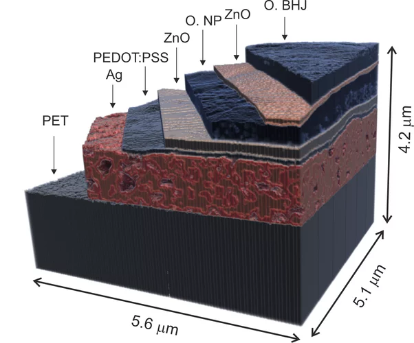
X-ray nanotomography aids the production of eco-friendly solar cells
Polymer solar cells are in the spotlight for sustainable energy production of the future. Characterization of these devices by X-ray nanotomography helps to improve their production using environmentally friendly materials.
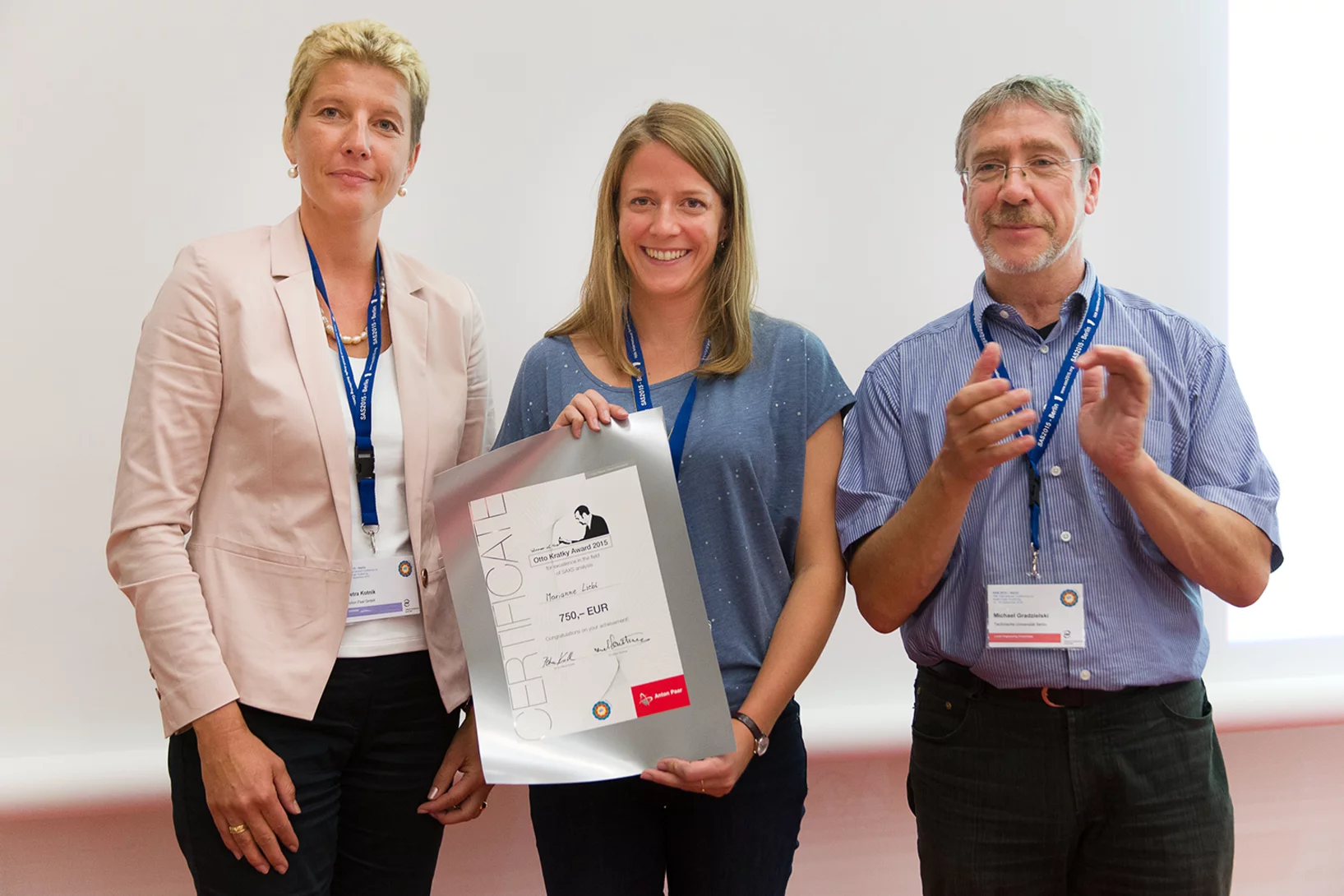
2015 Otto Kratky award
Marianne Liebi was awarded the 2015 Otto Kratky award by the Helmholtz-Centre Berlin for excellence in the field of small-angle X-ray scattering (SAXS) analysis. The award was bestowed in the last SAS2015 conference in Berlin. Marianne is a postdoctoral fellow in the coherent X-ray scattering group (CXS) in PSI, carrying out research in scanning SAXS measurement and analysis in 2D and 3D. Image credit ©HZB/Michael Setzpfandt
Element-Specific X-Ray Phase Tomography of 3D Structures at the Nanoscale
Recent advances in fabrication techniques to create mesoscopic 3D structures have led to significant developments in a variety of fields including biology, photonics, and magnetism. Further progress in these areas benefits from their full quantitative and structural characterization.
De l’intérieur d’une coquille d’œuf
La coquille d’un uf abrite de minuscules vésicules. Elles fournissent les substances qui stimulent et contrôlent la croissance de cette enveloppe solide. Grâce à une technique de tomographie novatrice, des chercheurs de l’Institut Paul Scherrer (PSI), de l’EPF Zurich et de l’Institut AMOLF aux Pays-Bas, ont réussi pour la première fois à obtenir une image en 3D de ces vésicules. Ils surmontent ainsi une limite à l’imagerie tomographique, et espèrent qu’un jour leur méthode profitera aussi à la médecine.
Multiresolution X-ray tomography, getting a clear view of the interior
Researchers at PSI have developed a technique that combines tomography measurements at different resolution levels to allow quantitative interpretation for nanoscale tomography on an interior region of interest of the sample. In collaboration with researchers of the institute AMOLF in the Netherlands and ETH Zurich in Switzerland they showcase their technique by studying the porous structure within a section of an avian eggshell. The detailed measurements of the interior of the sample allowed the researchers to quantify the ordering and distribution of an intricate network of pores within the shell.
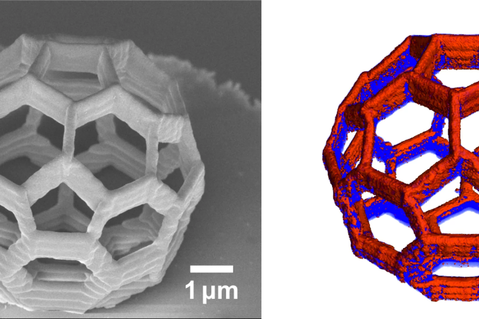
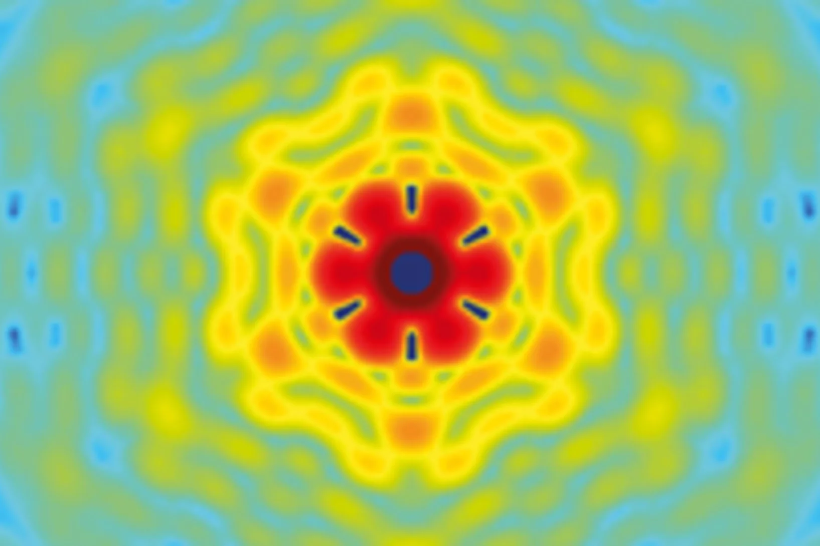
La 3D, au nanomètre près
Des chercheurs de l'Institut Paul Scherrer et de l'ETH Zurich ont créé des images en 3D de minuscules objets, et ont même réussi à visualiser au niveau de ces derniers des détails de 25 nanomètres (1 nanomètre = 1 million de millimètre). En plus de déterminer la forme de leurs objets d'étude, ils ont pu également mettre en évidence la façon dont un élément chimique donné (le cobalt) était réparti au sein de ces derniers, tout en étant capables d'établir si ce même élément était présent sous forme de liaison chimique ou sous forme pure.

Innovation Award on Synchrotron Radiation 2014 for high-resolution 3D hard X-ray microscopy
The 2014 Innovation Award on Synchrotron Radiation was bestowed to researchers Ana Diaz, Manuel Guizar-Sicairos, Mirko Holler, and Jörg Raabe from the Paul Scherrer Institut, Switzerland, for their contributions to method and instrumentation development, which have set new standards in high-resolution 3D hard X-ray microscopy.
Fast scanning coherent X-ray imaging using Eiger
The smaller pixel size, high frame rate, and high dynamic range of next-generation photon counting pixel detectors expedites measurements based on coherent diffractive imaging (CDI). The latter comprises methods that exploit the coherence of X-ray synchrotron sources to replace imaging optics by reconstruction algorithms. Researchers from the Paul Scherrer Institut have recently demonstrated fast CDI image acquisition above 25,000 resolution elements per second using an in-house developed Eiger detector. This rate is state of the art for diffractive imaging and even on a par with the fastest scanning X-ray transmission instruments. High image throughput is of crucial importance for both materials and biological sciences for studies with representative population sampling.
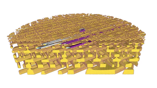
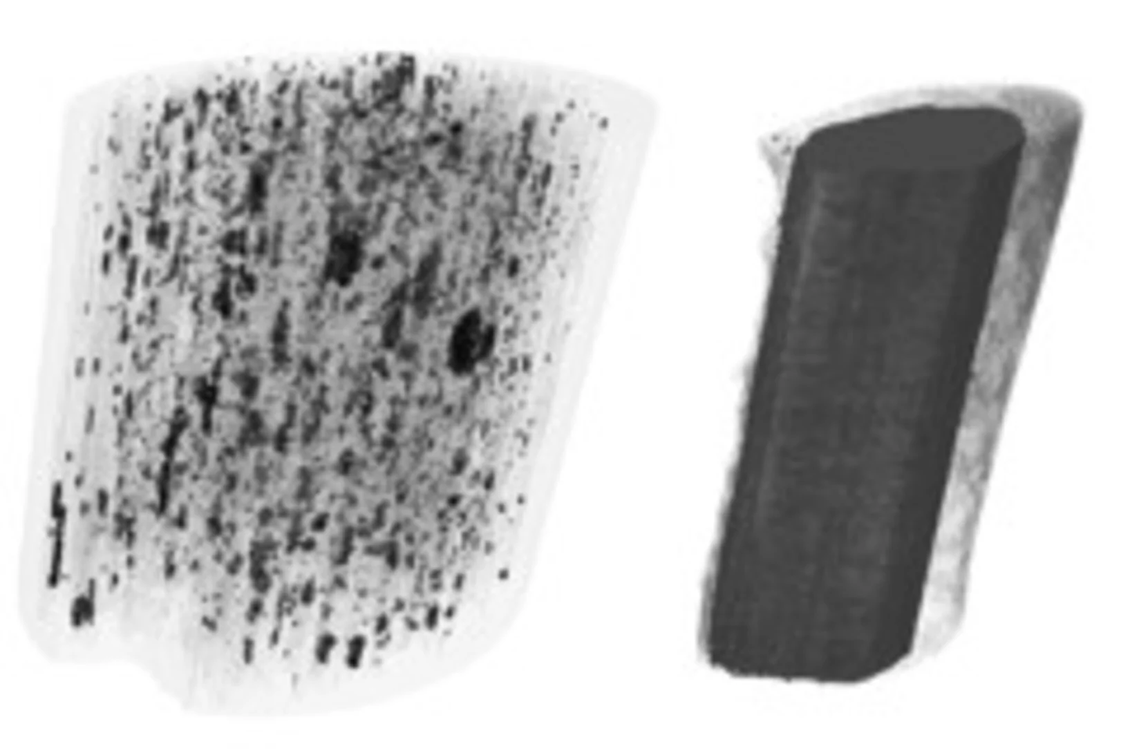
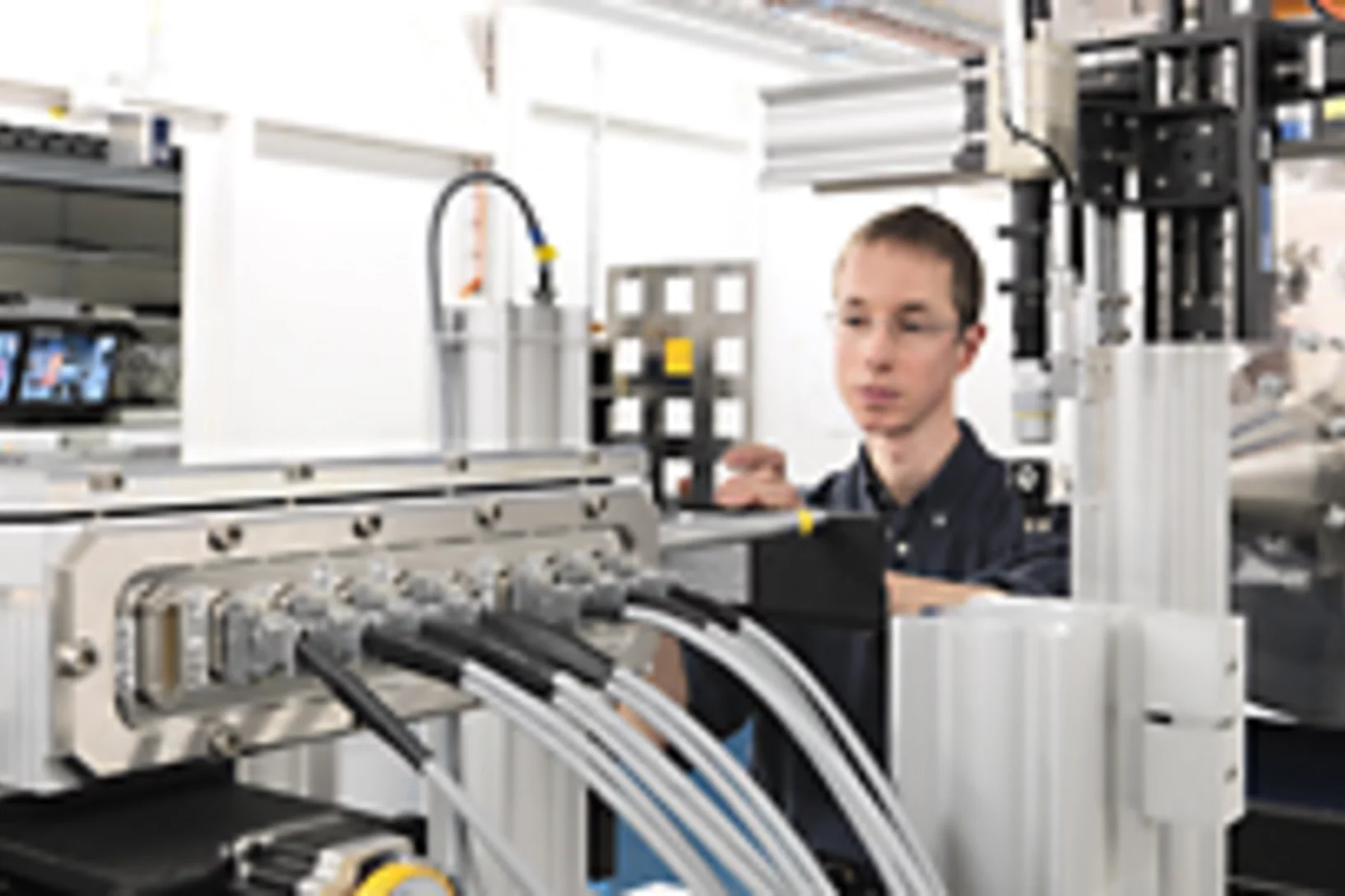
X-ray tomography reaches 16 nm isotropic 3D resolution
Researchers at PSI reported a demonstration of X-ray tomography with an unmatched isotropic 3D resolution of 16 nm in Scientific Reports. The measurement was performed at the cSAXS beamline at the Swiss Light Source using a prototype instrument of the OMNY (tOMography Nano crYo) project. Whereas this prototype measures at room temperature and atmospheric pressure, the OMNY system, to be commissioned later this year, will provide a cryogenic sample environment in ultra-high vacuum without compromising imaging capabilities. The researchers believe that such a combination of advanced imaging with state-of-the-art instrumentation is a promising path to fill the resolution gap between electron microscopy and X-ray imaging, also in case of radiation-sensitive materials such as polymer structures and biological systems.
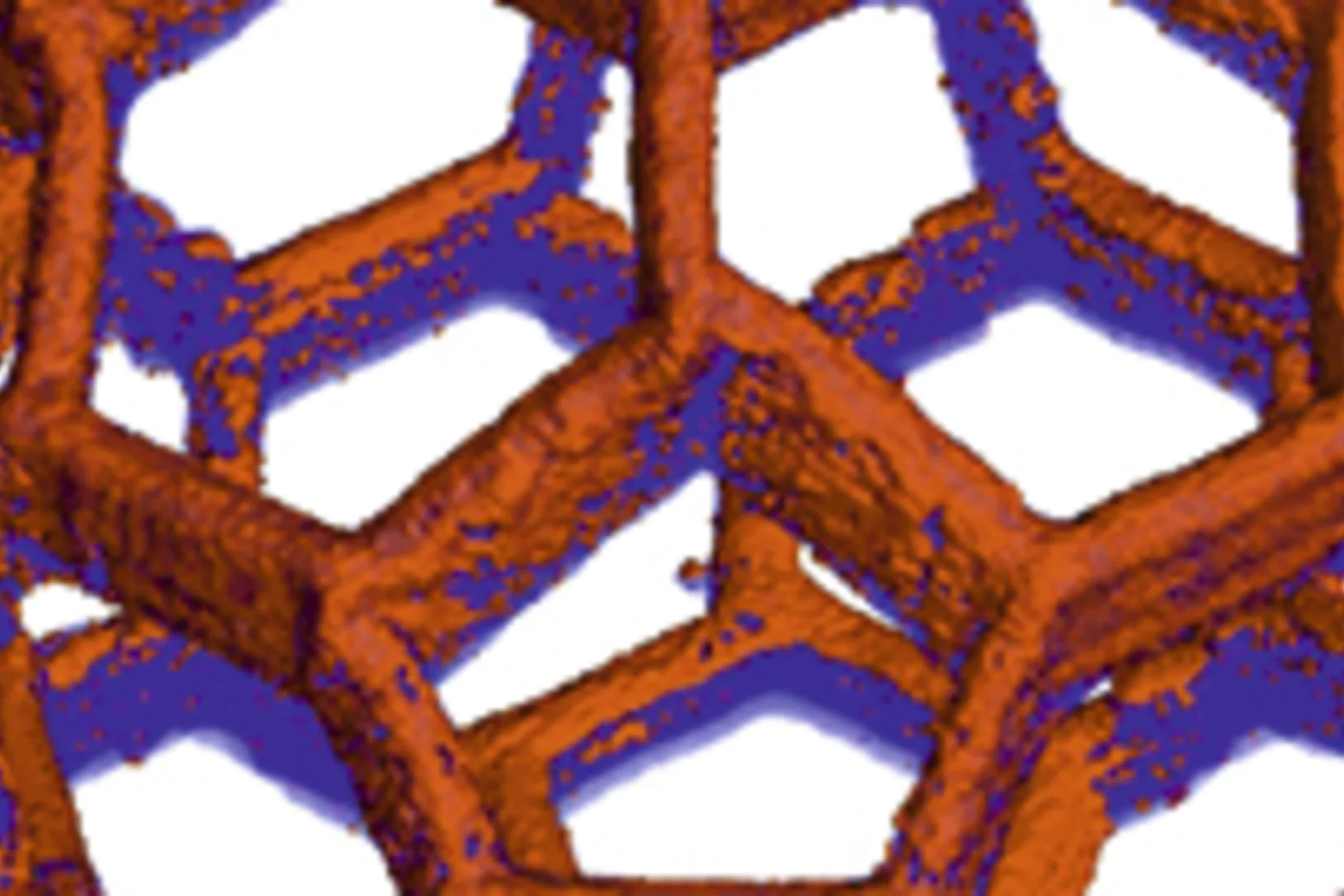
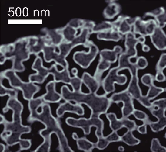
Unique insight into carbon fibers on the nanoscale
The investigation of the mechanical properties of carbon fibers benefits from highly resolved three-dimensional density maps within representative volumes, but such images are not easily obtained with standard methods. Scientists from the Paul Scherrer Institut in collaboration with Honda R&D in Germany have recently visualized density distributions on the sub-micrometer scale within entire carbon fiber sections, revealing surprising graphite distributions within the fibers. This capability will prove useful for the systematic characterization of fibers, contributing to the development of light and robust materials at lower costs.
Röntgen-Laser: Auf dem Weg zur Strukturbestimmung von Nanoteilchen
An Freie-Elektronen-Röntgen-Lasern wie dem zukünftigen SwissFEL des Paul Scherrer Instituts (PSI) sollen unter anderem die Strukturen von komplexen Nanoteilchen bis hin zu Biomolekülen untersucht werden. Dabei ist nicht nur die eigentliche Messung eine Herausforderung, sondern auch die Rekonstruktion der Struktur aus den Messdaten. Forscher des PSI haben nun einen optimierten mathematischen Weg aufgezeigt, wie man aus so gewonnen Messdaten eine deutlich bessere Auflösung bei der Bestimmung der Struktur eines einzelnen Teilchens erhält. Das Verfahren wurde an der Synchrotron Lichtquelle Schweiz des PSI erfolgreich getestet.Cette actualité n'existe qu'en allemand.
Imager des échantillons qui fluctuent à l'aide de rayons X
Les rayons X sont utilisés pour inspecter la structure à l'échelle nanométrique d'objets variés comme des cellules biologiques ou des dispositifs de stockage magnétiques. L'imagerie à très haute résolution impose cependant de très fortes contraintes, autant sur l'appareil que sur l'échantillon lui-même. Des chercheurs à la Technische Universität München et au PSI viennent de démontrer comment ces conditions peuvent être relaxées sans perte de qualité d'image. Ils ont de plus montré comment la même approche permet d'imager des échantillons qui fluctuent très rapidement, comme les matériaux magnétiques utilisés pour le stockage de données.
Nanoforscher untersuchen Karies
Forscher der Universität Basel und des Paul Scherrer Instituts konnten im Nanomassstab zeigen, wie sich Karies auf die menschlichen Zähne auswirkt. Ihre Studie eröffnet neue Perspektiven für die Behandlung von Zahnschäden, bei denen heute nur der Griff zum Bohrer bleibt. Die Forschungsergebnisse wurden in der Fachzeitschrift «Nanomedicine» veröffentlicht.Cette actualité n'existe qu'en allemand.
Röntgen-Methode hilft Hirnerkrankungen besser zu verstehen
Ein internationales Forschungsteam hat eine neue Methode entwickelt, mit der man detaillierte Röntgenbilder von Hirngewebe erstellen kann. Die Methode wurde verwendet, um die Myelinscheide der Nervenfasern sichtbar zu machen. Schäden an der Myelinscheide führen zu verschiedenen Erkrankungen wie etwa Multiple Sklerose. Die Anlage, an der diese Aufnahmen erstellt werden können, wird an der Synchrotron Lichtquelle Schweiz SLS des Schweizer Paul Scherrer Instituts betrieben.Cette actualité n'existe qu'en anglais et allemand.
Fortschritt für die Knochen-Forschung
Hochauflösendes Verfahren zur Nano-Computertomographie entwickeltEin neuartiges Nano-Tomographie-Verfahren, das von einem Team der TU München, des Paul Scherrer Instituts (PSI) und der ETH Zürich entwickelt wurde, erlaubt erstmals computertomographische Untersuchungen feinster Strukturen mit einer Auflösung im Nanometerbereich. Mit Hilfe der neuen Methode können etwa dreidimensionale Innenansichten fragiler Knochenstrukturen erstellt werden.Cette actualité n'existe qu'en anglais et allemand.