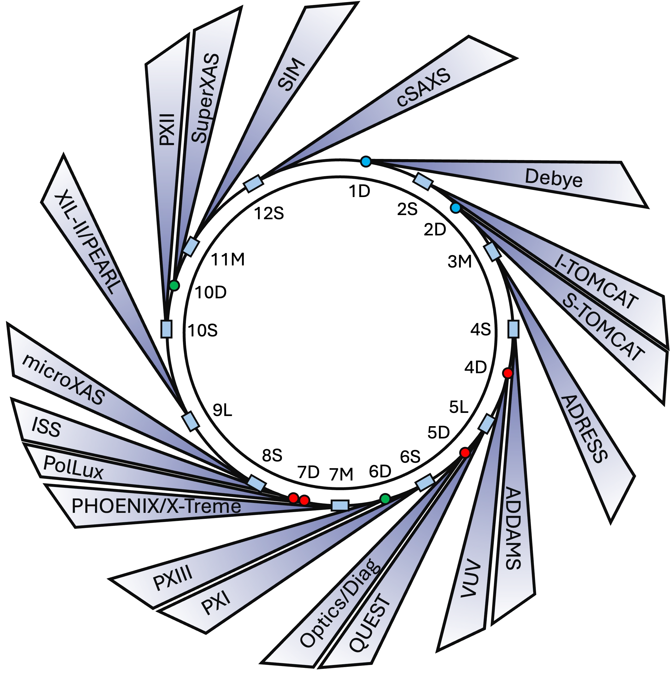
Beamline Finder
Show filters
| Long | Short | Full name Trier par ordre décroissant | Insertion Device |
|---|---|---|---|
| X04SA | ADDAMS | Advanced Diffraction for Material ScienceADDAMS (Advanced DiffrAction for Materials Science) is a beamline dedicated to powder diffraction and resonant X-ray diffraction within the hard X-ray range of 5 to 40 keV. The PD station operates a high-resolution powder diffractometer equipped with the novel Mythen III detector and a versatile table, using a frontal Pilatus 6M area detector. With its various attachments and in situ devices it allows investigations of samples in capillaries or bulk under a variety of conditions, including high throughput setups, ball-milling, gas pressure etc.. Time dependent phenomena can also be probed with powder diffraction using a local 22 kHz Eiger 1M. Surface diffraction is a unique tool for determining the detailed atomic structure of crystalline surfaces or the electronic and magnetic properties of materials. |
U14 |
| X03MA | ADRESS | Advanced Resonant SpectroscopiesThe beamline is constructed to deliver soft-X-ray radiation and has a Resonant Inelastic X-ray Scattering (RIXS) endstation and an Angle-Resolved Photoelectron Emission (ARPES) endstation. The scientific activity at the beamline is focussed on correlated systems (transition metals and rare earths) and nanostructures. |
UE44 |
| X03MA | ADRESS-SX-ARPES | Soft-X-ray Angle-Resolved Photoelectron SpectroscopyThe SX-ARPES facility is installed at the high-resolution undulator beamline ADRESS. The large probing depth and resonant photoexcitation achieved with soft X-rays allow investigations of k-resolved electronic structure of quantum materials including topological materials, buried interfaces, heterostructures and diluted impurity systems. The facility conducts extensive industrial research on materials for quantum computing. |
UE44 |
| X12SA | cSAXS | Coherent Small-Angle X-Ray ScatteringcSAXS is a beamline dedicated to high-resolution microscopy (using primarily ptychography, a coherent diffraction technique) and small-angle X-ray scattering (SAXS) within the hard X-ray range of 6 to ~30 keV. |
U17 |
| X01DA | Debye | X-ray absorption and X-ray diffraction (XAS and XRD)Debye is a beamline dedicated to sub-second high-throughput quasi-simultaneous X-ray absorption spectroscopy (quick XAS), X-ray diffraction (XRD) and total scattering (through the pair distribution function, PDF (available from 2027)). The beamline offers a tunable energy range from 4.5 to 60 keV, with a variable beamsize from 40 x 60 μm to 5 x 30 mm. Debye’s infrastructure includes: detectors for XAS (fast response ionization chambers, PIPS and a 7-element silicon drift detector SDD) and powder XRD (a Pilatus 6 M), a gas delivery system for reactive gases (up to 60 bars), several in situ and operando cells and a moveable sample environment table containing gas analytics (GC and MS), potentiostats and mass flow controllers for operando experiments. |
variable 3-5 Tesla bending magnet |
| X02SA | I-TOMCAT | I-TOMCAT: TOmographic Microscopy and Coherent rAdiology experimenTsThe I-TOMCAT beamline at the Swiss Light Source is a cutting-edge facility operated by the X-ray Tomography Group at PSI. It is designed to exploit the unique properties of synchrotron radiation for fast, non-destructive, and high-resolution 3D imaging across a wide range of scientific disciplines. I-TOMCAT is powered by a a U15 undulator (to be upgrade to a HTSU10 in 2027) and supports both absorption-based and phase-contrast imaging, with isotropic voxel sizes ranging from 50nm up to 0.65 microns and energy range of 8 to 50 keV (monochromatic). I-TOMCAT features three state-of-the-art endstations, dedicated to high-resolution, high-throughput, and dynamic tomographic imaging. |
U15 —> HTSU-10 |
| X07DB | ISS | In-situ spectroscopyAt the In Situ Spectroscopy beamline, ambient-pressure X-ray photoelectron spectroscopy (APXPS) and X-ray absorption spectroscopy (XAS) experiments can be performed. Two experimental chambers are available and can be connected to a shared electron analyzer, allowing the investigation of solid-gas and solid-liquid interfaces. |
Bending Magnet |
| X08SA | microXAS | microXAS - The Advanced Chemical Imaging BeamlineThe microXAS Chemical Imaging Beamline provides unique capabilities for Imaging Chemistry in Space and Time in a wide range of reactive systems at relevant spatial and temporal length scales. |
U17 |
| X03DA | PEARL | PhotoEmission and Atomic Resolution LaboratoryThe PEARL (PhotoEmission and Atomic Resolution Laboratory) beamline is dedicated to the structural, chemical and electronic characterisation of surfaces and adsorbates with atomic resolution. The beamline supports angle-resolved photoelectron spectroscopy (ARPES, XPS) and diffraction (XPD) and provides in-situ surface preparation and scanning tunnelling microscopy. |
Bending Magnet |
| X07MB | Phoenix | Photons for the Exploration of Nature by Imaging and XAFSThe PHOENIX (PHOtons for the Exploration of Nature by Imaging and XAFS) beamline is dedicated to X-ray Absorption (micro-) Spectroscopy (XAS) and imaging in both the soft and the rarely served tender X-ray’s covering the energyrange from 0.25 to 8 keV. both X-ray scanning microscopy (elemental mapping and chemical imaging) and micro-XAS can be performed with a spatial resolution of about 3 mm. The beamline covers important absorption edges of low-Z elements, in particular the K-edges of N-Fe and the L –edges of Ca-Er, providing unique research opportunities for material science, biology, energy research, environmental science, chemistry, catalysis, or cultural heritage. The beamline offers ample opportunity for in situ experiments (liquid cells, microjet, heating and cooling), which can be adapted to the user’s needs. Furthermore, a von Hamos spectrometer for emission spectroscopy in the energy range 2030-3100 eV and 3330-5060 eV is available. The beamline has two branchlines. The PHOENIX I branch uses a double crystal monochromator and covers energies from 1-8 keV, the soft x-ray branchline PHOENIX II invokes a planar grating monochromator and covers the range from 0.3-2 keV. |
UE38 |
| X07DA | PolLux | Highly Versatile Scanning Transmission X-Ray MicrospectroscopePolLux is a beamline dedicated to spectro-microscopy imaging at the soft X-ray (280-1500 eV) energy range. PolLux hosts a scanning transmission X-ray microscope that utilizes a diffractive Fresnel zoneplate lens to focus monochromatic X-ray onto a nanometric size (typical sizes between 10 and 20 nm) on an X-ray transparent sample. An image is obtained by scanning the sample with a piezoelectric stage and recording the transmitted intensity with a suitable point detector (either a photomultiplier tube or an avalanche photodiode). By having access to elemental contrast (through X-ray absorption spectroscopy), and several dichroic contrast mechanisms (X-ray circular and linear dichroism), both the elemental content of samples and their magnetic configurations can be resolved at the nanoscale. PolLux can also perform time-resolved measurements at the sub-ns timescales by means of pump-probe imaging, with a demonstrated temporal resolution of ca. 30 ps. Supporting RF electronics allow users to perform electrical pump-X-ray probe time-resolved investigations. |
Bending Magnet |
| X06SA | PXI | Macromolecular Crystallography (X06SA)X06SA (PXI) is the first macromolecular crystallography beamline at the Swiss Light Source. It is fully tunable from 5.7 to 17.5 keV with variable beam size from 5 um to 100 um, and is equipped with a flexible two-stage focusing X-ray optics system and a single–photon counting hybrid pixel area EIGER 16M (Dectris) detector. The PXI covers a wide range of MX applications from high-throughput crystallography to micro-crystallography and serial synchrotron crystallography. Room-temperaure and time-resolved MX are the latest features. The beamline team support both academic and proprietary users in on-site, remote, and fully-automated modes. |
U17 |
| X10SA | PXII | Macromolecular Crystallography (X10SA)The PXII beamline at the Swiss Light Source is a high-performance macromolecular crystallography facility primarily dedicated to proprietary users. It has recently undergone a significant upgrade, incorporating new optical components and enhanced automation. These developments will provide increased beam stability, higher flux, and variable beam sizes, while significantly streamlining experimental workflows. As a result, PXII is now even better suited for high-throughput data collection and the structure determination of challenging macromolecular samples. |
|
| X06DA | PXIII | Macromolecular Crystallography (X06DA)X06DA (PXIII) is a macromolecular crystallography beamline at the Swiss Light Source (SLS 2.0), available to both academic and proprietary users. PXIII receives synchrotron radiation from a 2.1 T superbend magnet. While it retains capabilities for low-energy applications, PXIII is primarily optimized for high-throughput data collection and unattended operation, under both cryogenic and room-temperature conditions. The current optical setup delivers a focused X-ray beam of approximately 30 × 20 µm² (h × v) at the sample position, with a photon flux of ~4 × 10¹¹ ph/s at 12.4 keV energy. The beam size can be adjusted between 10 × 10 µm² and 50 × 50 µm². The beamline is equipped with the new PILATUS4 2M Si detector, supporting data collection rates of up to 1.9 kHz. A state-of-the-art crystallization facility is in the immediate vicinity of the beamline, offering automated systems for both soluble and membrane protein crystallization, as well as support for fragment-based screening campaigns. |
Superbend |
| X02DA | S-TOMCAT | S-TOMCAT: TOmographic Microscopy and Coherent rAdiology experimenTsThe S-TOMCAT beamline at the Swiss Light Source is a cutting-edge facility operated by the X-ray Tomography Group at PSI. It is designed to exploit the unique properties of synchrotron radiation for fast, non-destructive, and high-resolution 3D imaging across a wide range of scientific disciplines. S-TOMCAT supports both absorption-based and phase-contrast imaging, with isotropic voxel sizes ranging from 0.16 to 11 micrometers and an energy range of 8 to 80 keV (monochromatic) as well as white beam from a powerful 5T superconducting magnet. Beam-size at sample can be as large as 50 mm horizontally. Phase contrast is achieved through propagation-based techniques, as well as grating interferometry, allowing researchers to visualise fine structural details even in weakly absorbing materials. |
5T superbend |
| X11MA | SIM | Surfaces / Interfaces MicroscopyThe permanent endstation of the SIM beamline is a Photoemission Electron microscope (PEEM) (Model: LEEM III, Elmitec GmbH). It allows to image samples using the photoelectric effect with very high spatial resolution. |
UE56 (twins) |
| X09LA | SIS-COPHEE | Surface and Interface Spectroscopy - Complete Photoemission Experiment (COPHEE)The Surface and Interface Spectroscopy beamline (SIS) provides a state-of-the-art experimental setup to study the electronic and atomic structure of surfaces. The beamline has been designed for high photon energy resolution with low harmonic contamination and flexible light polarization. The COPHEE endstation is dedicated to spin- and angle-resolved photoemission spectroscopy (SARPES), which can measure all properties of photoelectrons excited from a sample surface, namely energy, momentum and spin-polarization. The station reaches temperatures down to 17 K and offers an additional free contact for biasing or gating the sample. |
UE212 (twins) |
| X09LA | SIS-ULTRA | Surface and Interface Spectroscopy - Low-Temperature High-Resolution Angle-Resolved Photoemission (ULTRA)The Surface and Interface Spectroscopy beamline (SIS) provides a state-of-the-art experimental setup to study the electronic and atomic structure of surfaces. The beamline has been designed for high photon energy resolution with low harmonic contamination and flexible light polarization. ULTRA endstation is utilized for high-resolution angle-resolved photoemission spectroscopy (ARPES) at temperatures down to 4 K. |
UE212 (twins) |
| X10DA | Super-XAS | X-ray absorption and emission spectroscopy (XAS and XES)SuperXAS is a beamline dedicated to sub-second X-ray absorption spectroscopy (quick XAS), X-ray emission spectroscopy (XES) as well as laser pump- X-ray probe (XAS and XES) spectroscopy (100 ps to ms time resolution). The beamline offers a tunable energy range from 4.5 to 35 keV, with a variable beamsize (best focus 30 x 30 micro-meter). The infrastructure includes: detectors for XAS (fast response ionization chambers, PIPS and a 5-element silicon drift detector SDD), high resolution emission spectrometers (a 5-crystal conical crystal von Hamos spectrometer, a Johann spectrometer, a DuMond spectrometer (in commissioning)), a gas delivery system for reactive gases (up to 40 bars), several in situ and operando cells and a moveable sample environment table containing gas analytics (GC and MS), potentiostats and mass flow controllers for operando experiments. |
2.1 Tesla bending magnet |
| X04DB | VUV | Vacuum Ultraviolet RadiationThe VUV beamline, operated by the Reaction Dynamics Group, supplies bright, tunable vacuum ultraviolet bending-magnet synchrotron radiation from 5 to 30 eV (extendable to 100 eV, high harmonic-free between 5.5 and 21 eV). A grazing-incidence monochromator provides a resolving power of up to 10 000 with a photon flux of up to 1012 s−1 at 10 eV. The double-imaging photoelectron-photoion coincidence endstation (CRF-PEPICO) combines velocity-map imaging of electrons and ions with time-of-flight mass spectrometry of the latter. Isomer-specific photoion mass-selected photoelectron spectra identify stable reactants and products together with elusive intermediates, such as radicals, carbenes, and ketenes, in settings from low-pressure flames to catalytic or pyrolytic (micro)reactors and molecular beams. These reaction-level insights advance sustainable catalysis, combustion modelling, atmospheric chemistry, and astrochemical synthesis, while also refining our knowledge of molecular electronic structure, energetics, and thermochemistry. |
n/a |
| X07MA | X-Treme | X-Ray Magnetic Circular Dichroism under extreme ConditionsThe X-Treme beamline is dedicated to x-ray magnetic (circular or linear) dichroism technique in the soft x-ray range. The technique is element selective and is used for example for the study of magnetic anisotropy and exchange coupling. The energy range covers the L2,3-edges (2p to 3d transition) of 3d transition metals and M4,5-edges of lanthanides (3d to 4f transition), in addition to the K-edges of light elements like O, N, F. Scientific areas of interest are: single molecule magnets, magnetic nanocrystals, self-assembly of nanomagnets on surfaces and strongly correlated electron systems. |
UE54 |
| X09LB | XIL-II | X-Ray Interference Lithography and MetrologyThe XIL-II beamline has two branches. The lithography branch provides a spatially coherent beam in the Extreme Ultraviolet (EUV) energy range (typically around 92 eV) and is equipped with an interference lithography end station (EUV-IL) used for photoresist characterization and for the manufacturing of periodic nanostructures with half pitch as small as 5 nm. The metrology branch is equipped with an SG monochromator providing a coherent beam with tunable wavelength and a λ/dλ ratio of 1500 at 13.5 nm. Its end station is REGINE, an actinic lensless microscope dedicated to EUV mask, pellicle and wafer metrology with a maximum resolution of 34 nm. |
U70 |
1) Insertion Device
The highest brightness at a beamline can be reached by means of insertion devices (Undulators, marked with U). The number behind the letter for the device is the period length in mm. The letter E in the name of an undulator means elliptical polarization is possible.
2) Bending magnet beamlines
This type of beamline uses the radiation from one of the bending magnets of the 7-bend achromats of SLS 2.0