
News & Scientific Highlights

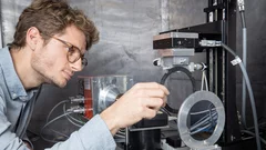
3D printing silica aerogels at the micrometer scale
A group of EMPA and ETH Zürich researchers have developed a new method to directly write ink made of silica aerogels in 3D. Thanks to X-ray phase contrast tomography at the TOMCAT beamline they characterized the resulting printed material with different compositions. Their results were published in Nature on August 18, 2020.
")
Phase contrast microtomography reveals nanoparticle accumulation in zebrafish
Metal-based nanoparticles are a promising tool in medicine – as a contrast agent, transporter of active substances, or to thermally kill tumor cells. Up to now, it has been hardly possible to study their distribution inside an organism. Researchers at the University of Basel in collaboration with the TOMCAT team have used phase contrast X-ray tomographic microscopy to take high-resolution captures of the nanoparticle aggregation inside zebrafish embryos.
The study was published in the journal Small and featured on the cover of its current issue.
4 times compression factor for tomographic data feasible
In a recent study, TOMCAT has shown that lossy compression by a factor of at least 3 to 4 of raw acquisitions generally does not affect the reconstruction quality and that higher factors (six to eight times) can be achieved for tomographic volumes with a high signal-to-noise ratio as it is the case for phase-retrieved datasets. This finding is relevant to current challenges on large tomography data management and storage especially at synchrotron facilities. The results of this study was published in Journal of Synchrotron Radiation.
Miniaturized fluidic circuitry observed in 3D
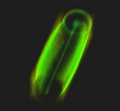
The team of Prof. Thomas Hermans at the University of Strasbourg in France managed to create wall-less aqueous liquid channels called anti-tubes. Thanks to X-ray phase contrast tomography at the TOMCAT beamline those anti-tubes could be observed in 3D. The exciting results were published in Nature on May 6, 2020.

Rapid 3D directional small-angle scattering imaging achieved at TOMCAT
Researchers from the TOMCAT beamline have developed a small-angle scattering tensor tomography method to visualize microscopic features within a macroscopic field of view with unprecedented data acquisition speed. The results of the study were published in Applied Physics Letters on April 1, 2020.

X-ray Imaging for Biomedicine: Imaging Large Volumes of Fresh Tissue at High Resolution
The TOMCAT beamline at the Swiss Light Source specializes in rapid high-resolution 3-dimensional tomographic microscopy measurements with a strong focus on biomedical imaging. The team has recently developed a technique to acquire micrometer-scale resolution datasets on the entire lung structure of a juvenile rat in its fresh natural state within the animal’s body and without the need for any fixation, staining or other alteration that would affect the observed structure (E. Borisova et al., 2020, Histochem Cell Biol).

Towards dynamic feedback control during time-resolved CT at TOMCAT
Researchers from the CWI in Amsterdam and the TOMCAT beamline have developed and implemented a real-time CT reconstruction, visualisation, and on-the-fly analysis approach to monitor dynamic processes as they occur. With processes of multiple sets of CT slices per second, this represents the next crucial step towards adaptive feedback control of time-resolved in situ tomographic experiments. The results of this study were published in Scientific Reports on December 5, 2019.
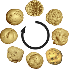
Animal embryos evolved before animals
Detailed characterization of cellular structure and development of exceptionally preserved ancient tiny fossils from South China by synchrotron based X-ray tomographic microscopy at TOMCAT led an international team of researchers from the University of Bristol and Nanjing Institute of Geology and Palaeontology to the discovery that animal-like embryos evolved long before the first animals appear in the fossil record.
Radiographier rapidement et précisément des matériaux composites renforcés de fibres
Des chercheurs de l’Institut Paul Scherrer PSI ont mis au point une méthode de diffusion des rayons X aux petits angles qui peut être utilisée pour le développement ou le contrôle qualité de matériaux composites novateurs renforcés de fibres. Grâce à elle, les analyses de ces matériaux pourraient se faire à l’avenir non seulement par recours aux rayons X issus de sources puissantes comme la Source de Lumière Suisse SLS, mais aussi avec le rayonnement issu de tubes à rayons X conventionnels.