
Lab News & Scientific Highlights
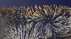
Plongée dans un aimant
Pour la première fois, des chercheurs ont réussi à visualiser les directions de l'aimantation dans un objet magnétique tridimensionnel. Les plus petits détails de leur visualisation mesuraient moins d'un dixième de millième de millimètre. Un type de motif exceptionnel est ressorti dans la structure qu'ils ont fait apparaître: des singularités magnétiques appelées points de Bloch, jusque-là connues uniquement en théorie.
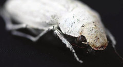
Photonic structure of white beetle wing scales: optimized by evolution
A very thin layer on this beetle’s wings exhibits a complicated structure on the nanoscale that gives them a bright white color. X-ray nanotomography acquired at the Swiss Light Source provides a faithful image of this structure in three dimensions with which scientists can confirm its evolutionary optimization: just enough material for an efficient reflection of white light.
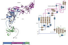
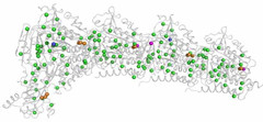
Towards understanding of human betacoronavirus HKU1 life cycle
Researchers from China and USA join forces with Swiss Light Source (SLS) macromolecular crystallography (MX) beamline scientists in a study, which aims at understanding an important step in the life cycle of the human betacoronavirus HKU1.
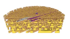
La radiographie en 3D permet de visualiser les moindres détails d’une puce informatique
Des chercheurs de l’Institut Paul Scherrer PSI ont réalisé des radiographies détaillées en 3D d’une puce informatique usuelle. Dans le cadre de leur expérience, ils ont analysé une petite portion de puce qu’ils avaient préalablement découpée. Durant la mesure, cet échantillon est resté intact. Pour les fabricants, déterminer si la structure de leurs puces est conforme aux normes représente un défi. Ces résultats constituent donc une possibilité d’application importante pour un procédé spécifique de tomographie à rayons X que les chercheurs du PSI développent depuis quelques années.
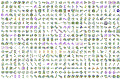
1000 Structures solved at X06DA-PXIII
The macromolecular crystallography beamline X06DA-PXIII has reached 1,000 structures in the Protein Data Bank (PDB) on February 22, 2017.
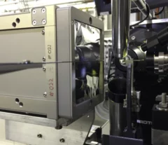
First protein structure solved using the JUNGFRAU detector!
JUNGFRAU is a charge-integrating, two-dimensional pixel detector developed at the Paul Scherrer Institut for use at free-electron lasers, in particular SwissFEL, and synchrotron light sources. On the 10th October, the first protein crystallography experiment using the JUNGFRAU detector, was performed at the beamline X06SA (PXI) of the Swiss Light Source by the members of the Protein Crystallography and Detectors groups at PSI.
Call for expressions of interest: Beamline partners at the SLS for PX II and PX III
We invite companies and institutions to secure access to the beamlines X10SA/PX II and X06DA/PX III through a long term contract.

Single shot grating interferometry demonstrated using direct conversion detection
Researchers at the Paul Scherrer Institute's Swiss Light Source in Villigen, Switzerland, have developed an X-ray grating interferometry setup which does not require an analyzer grating, by directly detecting the fringes generated by the phase grating with a high resolution detector. The 25um pitch GOTTHARD microstrip detector utilizes a direct conversion sensor in which the charge generated from a single absorbed photon is collected by more than one channel. Therefore it is possible to interpolate to achieve a position resolution finer than the strip pitch.
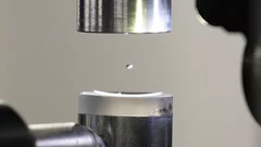
Expérience dans une goutte en lévitation
La structure exacte des protéines est normalement déterminée au PSI par la technique de diffraction des rayons X. Deux scientifiques du PSI viennent de l’améliorer de façon astucieuse: au lieu d’immobiliser les protéines, ils les ont étudiées dans une goutte de liquide en lévitation.